
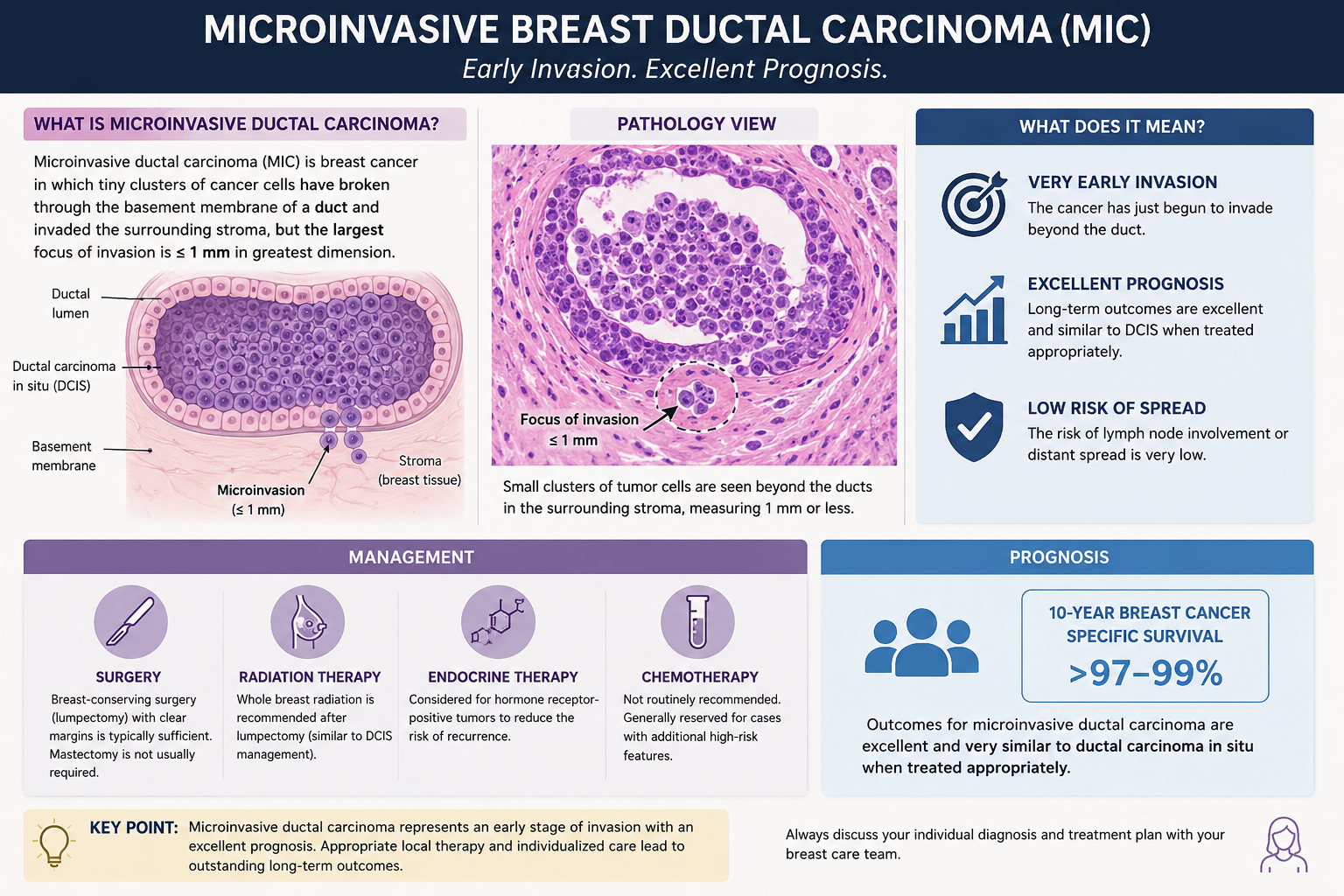
A breast cancer report can feel frightening. microinvasive breast carcinoma means very small invasion; learn what to ask next.
microinvasive breast carcinoma means that breast cancer cells have just started to break out of a pre-invasive area and invade nearby breast tissue, but only in a very tiny amount. In pathology, this finding is usually linked to ductal carcinoma in situ, often shortened to DCIS, with one or more microscopic foci of invasion. The word “carcinoma” can sound overwhelming, but the word “microinvasive” carries very specific meaning about how small the invasive component is.
Receiving a breast pathology report with unfamiliar terms can make time feel as if it stops. Many patients see the phrase microinvasive breast carcinoma and immediately wonder whether this means a more dangerous cancer, whether treatment must change, or whether something was missed earlier. Clear information cannot remove every worry, but it can make the next conversation with the care team more grounded and less frightening.
A pathology diagnosis is not just a label. It is a map that helps surgeons, oncologists, radiation oncologists, and patients decide what should happen next. For a broader overview of how reports are organized, patients may find What Is a Pathology Report? helpful alongside the specific explanation below.
Microinvasive Breast Carcinoma — What It Actually Means
Microinvasive breast carcinoma is diagnosed when invasive cancer measures no more than 1 millimeter in greatest dimension. A useful analogy is a small crack in a wall: Most of the process may still be on one side of the wall, but a tiny focus has just crossed through. In breast pathology, that “wall” is the basement membrane, a thin natural boundary that separates ducts from surrounding breast tissue. When cancer cells cross the basement membrane, even by a very small amount, the diagnosis changes from purely in situ disease to microinvasive disease.
This finding most often appears in a breast pathology report that also describes ductal carcinoma in situ. Ductal carcinoma in situ means abnormal cancer cells are present inside the breast ducts but have not broadly invaded the surrounding tissue. Microinvasion means pathologists found a very small area where cells moved beyond the duct boundary. The invasive focus is measured carefully under the microscope, and tumor size becomes central because the cutoff is so small.
Pathologists often examine multiple tissue sections to confirm microinvasive breast carcinoma because the difference between DCIS and invasion can be subtle. Special stains may be used to highlight the myoepithelial cell layer, which normally outlines breast ducts and supports the interpretation of whether the basement membrane has been crossed. Hormone receptors, HER2 testing, grade, margins, and other features may also appear in the report. For patients trying to understand where each item fits, Understanding Your Pathology Report: How to Read It with Confidence can make the layout feel less mysterious.
Why Your Report Shows This Finding
A report may show microinvasive breast carcinoma because a biopsy or surgical specimen found mostly ductal carcinoma in situ with a tiny invasive area. Ductal carcinoma in situ can grow along the duct system and sometimes develop features that raise concern for possible invasion, such as high nuclear grade, comedo necrosis, or a mass-like area on imaging. The pathologist samples the tissue, looks at the architecture, and searches for areas where cells have crossed the basement membrane. If invasion is present but measures 1 millimeter or less, the term microinvasive is used.
The diagnosis can be challenging because breast tissue is three-dimensional, but microscope slides show very thin two-dimensional slices. A tiny group of cells may appear suspicious in one section and clearer in another. Pathologists look for the loss of the myoepithelial layer, changes in the surrounding stroma, and the relationship of the cells to ducts and lobules. Immunohistochemistry can help by staining myoepithelial markers, although the final diagnosis still depends on the full pattern rather than one stain alone.
A breast pathology report may also include tumor size, margin status, hormone receptors, and HER2 results because these details help determine management. Tumor size can be confusing when there is extensive ductal carcinoma in situ but only a tiny invasive component. For staging, the invasive component matters most, while the extent of ductal carcinoma in situ can affect surgical planning and margins. Hormone receptors help identify whether endocrine therapy may be useful, and HER2 status may be assessed depending on the invasive focus and local practice.
How Serious Is Microinvasive Breast Carcinoma?
Microinvasive breast carcinoma is serious because it is a form of invasive breast cancer, but it is usually at the very earliest end of the invasive spectrum. The word “invasive” understandably causes fear, yet the word “microinvasive” means the invasive component is extremely small. Many patients with this diagnosis do very well, especially when the disease is completely removed and the lymph nodes are negative. The seriousness depends on the full breast pathology report, not on one phrase alone.
Microinvasive breast carcinoma is commonly staged as T1mi when the invasive focus is 1 millimeter or smaller. This is different from larger invasive breast cancer, where tumor size may be measured in millimeters or centimeters beyond that cutoff. The presence of extensive ductal carcinoma in situ can still matter because it may influence whether additional surgery is needed to achieve clear margins. However, the tiny invasive component is a key reason the prognosis is often favorable compared with larger invasive breast cancer.
The main questions are whether the margins are clear, whether there is more than one microinvasive focus, whether a lymph node biopsy was performed, and whether any lymph nodes contain cancer. A lymph node biopsy may be recommended in some cases, particularly when mastectomy is planned or when the invasive component is confirmed. Most patients with microinvasive disease do not have lymph node involvement, but checking the nodes can provide important reassurance and staging information. Hormone receptors and HER2 results also help the oncology team decide whether treatments beyond surgery and radiation should be discussed.

What Happens Next: Treatment And Monitoring
After microinvasive breast carcinoma is diagnosed, the next steps usually depend on the type of procedure already performed. If the finding came from a core needle biopsy, surgery is typically needed to remove the area more completely and evaluate the extent of disease. If the diagnosis came from lumpectomy or mastectomy tissue, the care team reviews margins, tumor size, ductal carcinoma in situ extent, and lymph node biopsy results. The breast pathology report becomes the starting point for a treatment plan rather than a final decision by itself.
Treatment may include lumpectomy followed by radiation, mastectomy in selected cases, sentinel lymph node biopsy, endocrine therapy if hormone receptors are positive, or additional evaluation if HER2 is positive. Not every patient needs every treatment. The distinction between microinvasive disease and larger invasive breast cancer can affect how aggressive the recommendations are. Decisions are usually made in a multidisciplinary setting where pathology, imaging, surgery, medical oncology, and radiation oncology are considered together.
Monitoring after treatment is usually individualized. Follow-up may include breast exams, mammography, review of any new symptoms, and long-term discussion of risk reduction. Patients may also hear about Ki-67, although this marker is not always central in microinvasive disease; when it appears, What Does Ki-67 Mean and How High Is Concerning? may help explain its role. The most useful follow-up plan is one that matches the final diagnosis, the surgery performed, and the patient’s overall health.
Questions To Ask Your Doctor Or Pathologist
The most helpful questions are specific to the wording of the report. A patient does not need to memorize pathology terminology before the appointment. The goal is to understand which part of the cancer is in situ, which part is invasive, and what information still needs confirmation.
- Was the microinvasive breast carcinoma measured as 1 millimeter or less?
- Is there one focus of microinvasion, or are there multiple foci?
- How extensive is the ductal carcinoma in situ around the microinvasive area?
- Are the surgical margins clear for both the invasive component and ductal carcinoma in situ?
- Was a lymph node biopsy done, and were any lymph nodes positive?
- What are the hormone receptors and HER2 results, and were they tested on the invasive focus?
- Does the breast pathology report need additional stains, review, or clarification before treatment is finalized?
A second pathology opinion can be valuable when the distinction between ductal carcinoma in situ, microinvasion, and larger invasive breast cancer is unclear. This does not mean the original pathologist made an error; it means small findings sometimes deserve another expert review before major treatment decisions. Patients considering this step may find When Should You Get a Second Pathology Opinion? useful.
Expert review is especially reasonable when the report mentions suspicious invasion, possible microinvasion, close margins, unusual biomarker results, or disagreement between imaging and pathology. A second review typically examines the original slides, any immunohistochemistry, and the final report language. Clearer wording can help the care team choose treatment with more confidence.
Frequently Asked Questions
Is microinvasive breast carcinoma the same as breast cancer?
Yes. Microinvasive breast carcinoma is considered invasive breast cancer, but the invasive component is extremely small. It is usually found with ductal carcinoma in situ and is often staged as T1mi when the invasion is 1 millimeter or less. This diagnosis should be taken seriously, but it does not carry the same meaning as a much larger invasive tumor.
Can microinvasive breast carcinoma spread to lymph nodes?
Microinvasive breast carcinoma can spread to lymph nodes, but this is uncommon compared with larger invasive breast cancer. A lymph node biopsy may be recommended depending on the type of surgery, imaging findings, and institutional practice. Negative lymph nodes are reassuring and help confirm early-stage disease. The care team uses the node result together with tumor size, margins, and biomarkers.
Does microinvasion mean chemotherapy is needed?
Microinvasion does not automatically mean chemotherapy is needed. Many patients with microinvasive disease do not receive chemotherapy, especially when lymph nodes are negative and the invasive focus is tiny. Hormone receptors, HER2 status, grade, and the overall clinical picture can influence recommendations. A medical oncologist can explain whether systemic treatment is likely to provide meaningful benefit.
What does T1mi mean on a breast pathology report?
T1mi is a staging term used when the invasive breast cancer component measures 1 millimeter or less. It is commonly used for microinvasive breast carcinoma. The “T” describes the primary tumor, and “mi” stands for microinvasion. This staging term should be interpreted alongside lymph node status and whether there is associated ductal carcinoma in situ.
Should I get a second opinion for microinvasive breast carcinoma?
A second opinion can be helpful when microinvasive breast carcinoma affects choices about surgery, radiation, lymph node biopsy, or medication. It may also help if the report language is uncertain, such as “suspicious for microinvasion” or “cannot exclude invasion.” The review usually focuses on whether the basement membrane has truly been crossed and whether the invasive focus is accurately measured. Practical steps are outlined in How to Get a Second Opinion on Your Pathology Diagnosis.
Microinvasive breast carcinoma is a frightening phrase, but it describes a very specific and very small amount of invasion. The safest path is to understand the complete report, confirm the key measurements and margins, and ask whether expert pathology review would change any treatment decision. Honest Pathology consultations can help patients and families translate complex breast pathology language into clear questions for the treating team.
References:
National Cancer Institute — Breast Cancer Treatment (PDQ®) Patient Version
National Cancer Institute — Pathology Reports
National Cancer Institute Dictionary of Cancer Terms — Carcinoma In Situ
