Worried by iron on a liver biopsy? hepatic iron grading helps explain the pattern, amount, and next questions to ask.
hepatic iron grading is the way pathologists describe how much iron is seen in liver tissue and where that iron is located. This information usually comes from a liver biopsy examined under the microscope, often with a special iron stain. The grade does not diagnose every cause by itself, but it gives the treating doctor important clues about iron overload, liver injury, and whether additional blood or genetic testing may be needed.
Seeing the word “iron” in a biopsy result can feel unsettling, especially when the report also mentions scarring, inflammation, or chronic liver disease. Many patients wonder whether this means hemochromatosis, cirrhosis, cancer, or something urgent. In many cases, the finding is only one piece of a larger clinical picture.
A pathology report is like a carefully marked map rather than a final travel itinerary. It shows where the iron is, how much is present, and what else is happening in the tissue. For patients trying to understand the report line by line, a plain-language overview such as Understanding Your Pathology Report can make the wording less intimidating.
Hepatic Iron Grading — What It Actually Means
Hepatic Iron Grading describes the amount of stainable iron in a liver biopsy, usually on a scale from absent to marked. Different laboratories may use slightly different scoring systems, but many reports use a 0 to 4 scale. Grade 0 means no visible iron, while higher grades mean progressively more iron is present in the tissue. The phrase hepatic iron grading does not mean the liver is being “graded” like a cancer stage; it means the iron deposits are being semi-quantified under the microscope.
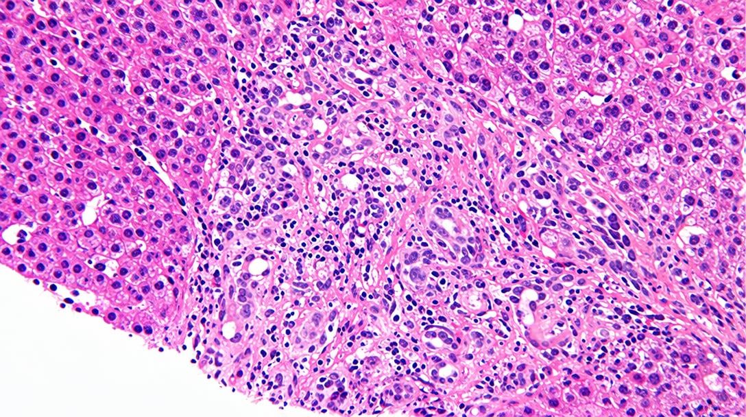
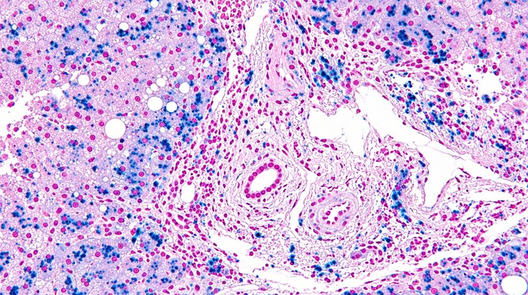
The most common method uses an iron stain, often called Prussian blue, which makes iron deposits appear blue within the liver tissue. Without the iron stain, small deposits may be difficult or impossible to appreciate on the routine hematoxylin and eosin slide. A helpful analogy is using a highlighter on a printed page: the words were already there, but the special stain makes one feature stand out. In the same way, the iron stain helps the pathologist see the pattern and amount of iron more clearly.
The location of iron matters as much as the amount. Iron may be concentrated inside liver cells, known as hepatocytes, or in scavenger-type cells called Kupffer cells. A liver biopsy with mostly hepatocellular iron can suggest one set of possibilities, while iron mainly in Kupffer cells can suggest another. The pathology report may describe both the grade and the distribution because hepatic iron grading is most useful when paired with the pattern.
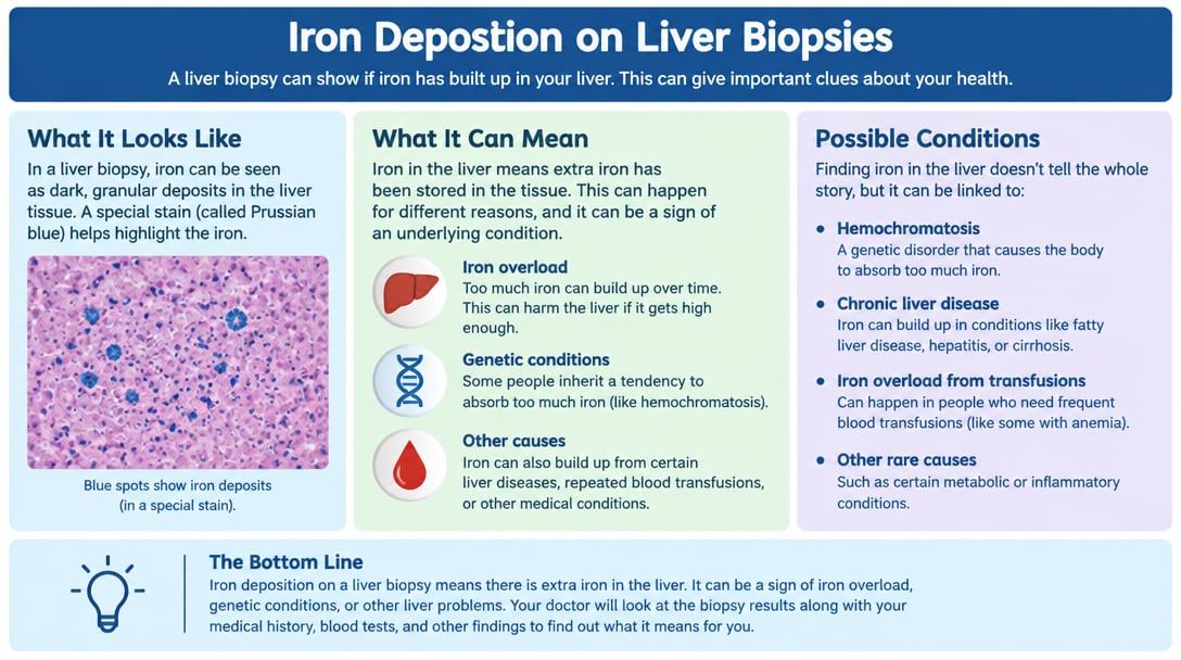
Why Your Report Shows This Finding
A report may show iron deposition for several reasons, and not all of them mean inherited hemochromatosis. Iron overload can occur when the body absorbs too much iron, receives repeated blood transfusions, has chronic liver disease, or has certain blood disorders. Alcohol-related liver disease, fatty liver disease, viral hepatitis, and long-standing inflammation can also be associated with increased iron in some cases. The liver biopsy gives a tissue-level view, but the treating clinician must connect that view with blood tests and the patient’s history.
Pathologists look for the intensity of Prussian blue staining and the exact cells that contain the pigment. Hepatocellular iron often draws attention to disorders of iron absorption and storage, including hereditary hemochromatosis. Iron in Kupffer cells may be seen after transfusions, with increased breakdown of red blood cells, or in some inflammatory settings. Mixed patterns can occur, which is why hepatic iron grading is usually interpreted alongside ferritin, transferrin saturation, liver enzymes, and clinical context.
The wording in a pathology report may include terms such as “mild iron deposition,” “2+ iron,” “grade 3 of 4,” or “iron present in hepatocytes and Kupffer cells.” These phrases can sound precise but still require explanation. A report answers what is visible in the biopsy sample, not every reason why it happened. For patients learning the structure of a report, What Is a Pathology Report? explains how microscopic descriptions, diagnoses, and comments fit together.
How Serious Is Hepatic Iron Grading on a Liver Biopsy?
Hepatic Iron Grading becomes more serious when the grade is high, the iron is mainly within hepatocytes, or the biopsy also shows significant fibrosis or cirrhosis. Mild iron alone may not mean severe disease, especially if blood tests are only slightly abnormal. A small amount of iron can be seen in several liver conditions and may not change treatment by itself. The level of concern depends on the whole picture, including symptoms, family history, ferritin level, transferrin saturation, and other liver findings.
In practical terms, hepatic iron grading helps separate “a small amount of iron was noticed” from “there is enough iron to raise concern for clinically meaningful iron overload.” Marked hepatocellular iron often prompts evaluation for hereditary hemochromatosis, especially when transferrin saturation is high. Marked iron in Kupffer cells may lead the clinical team to review transfusion history, hemolysis, or other causes of secondary iron overload. The biopsy does not replace blood testing, but it can confirm that iron is truly being stored in liver tissue.
The grade also matters because long-standing iron overload can injure the liver and contribute to scarring. When iron is accompanied by bridging fibrosis or cirrhosis, doctors often treat the result with more urgency. When iron is mild and fibrosis is absent, monitoring and targeted testing may be enough. This is why the same phrase, hepatic iron grading, can carry different meanings in different patients.
What Happens Next: Treatment and Monitoring
After a liver biopsy shows iron deposition, the next step is usually correlation with blood tests. Common tests include ferritin, transferrin saturation, complete blood count, liver enzymes, and sometimes genetic testing for HFE-related hemochromatosis. If iron overload appears likely, treatment may include therapeutic phlebotomy, changes in iron supplementation, or management of an underlying blood or liver condition. The exact plan depends on why the iron is present, not just the iron stain result.
Monitoring may include repeat blood work rather than another biopsy. Ferritin and transferrin saturation are often followed over time to see whether iron levels are improving. If there is significant fibrosis, the medical team may also monitor for complications of chronic liver disease. A liver biopsy can show scarring, inflammation, fat, and iron in the same tissue sample, which is why it can be so useful when several conditions are possible.
Patients may also see comments about hepatocellular iron, Kupffer cells, or Prussian blue in follow-up notes. These terms are not meant to frighten patients; they are the technical language used to communicate a tissue pattern. If the diagnosis seems uncertain or if treatment decisions are major, a second review of the slides may be reasonable. Guidance on timing and reasons for review is available in When Should You Get a Second Pathology Opinion?.
Questions to Ask Your Doctor or Pathologist
Clear questions can turn an intimidating pathology report into a focused conversation. For hepatic iron grading, the most helpful questions ask about amount, pattern, cause, and next steps. Patients may want to bring both the biopsy report and recent blood test results to the visit. The following questions are often useful:
- What grade of iron was reported, and what grading scale did the laboratory use?
- Was the iron mainly in hepatocytes, Kupffer cells, or both?
- Did the iron stain show mild, moderate, or marked iron deposition?
- Does the pattern suggest hereditary hemochromatosis or secondary iron overload?
- How do the biopsy findings compare with ferritin and transferrin saturation results?
- Did the liver biopsy also show fibrosis, cirrhosis, steatosis, or active hepatitis?
- Would a second pathology review change the diagnosis or treatment plan?
A pathologist can often clarify whether the grade is straightforward or whether the pattern is mixed. Some reports include a diagnostic comment, while others only list the microscopic finding. When the wording is brief, direct questions can uncover how confident the interpretation is.
A second opinion is not a sign that the first report was wrong. It is a careful rereading of the same tissue by another qualified pathologist, sometimes with additional stains or clinical information. For patients considering this step, How to Get a Second Opinion on Your Pathology Diagnosis explains the process in practical terms.
Frequently Asked Questions
What does iron in a liver biopsy mean?
Iron in a liver biopsy means that stainable iron is visible in the sampled liver tissue. The finding may reflect hereditary hemochromatosis, secondary iron overload, chronic liver disease, transfusion history, or other clinical factors. The meaning depends on the grade, the pattern, and the blood test results. Hepatic iron grading helps organize this information so the clinical team can decide what to evaluate next.
Is hepatic iron grading the same as hemochromatosis?
Hepatic iron grading is not the same as a diagnosis of hemochromatosis. It describes the amount and distribution of iron seen under the microscope. Hemochromatosis is one possible cause, especially when hepatocellular iron is prominent and transferrin saturation is high. Additional clinical, laboratory, and sometimes genetic information is needed before that diagnosis is made.
What is a normal hepatic iron grade?
Many liver biopsy reports describe normal iron as absent or grade 0, although exact terminology varies by laboratory. A very small amount may be described as trace or mild in some settings. The pathologist usually interprets the grade in relation to the rest of the biopsy. A normal or near-normal grade is generally less concerning than moderate or marked iron deposition.
Can iron overload damage the liver?
Yes, persistent iron overload can injure liver cells and contribute to fibrosis over time. The risk is higher when iron levels are substantial and present for many years. A liver biopsy can show whether scarring has already developed. Treatment and monitoring can reduce risk when the cause of iron overload is identified early.
Can hepatic iron grading change after treatment?
Hepatic iron grading can improve after effective treatment, but repeat biopsy is not always needed to prove that improvement. Doctors often monitor ferritin and transferrin saturation instead. If the original liver biopsy showed significant fibrosis or an unclear pattern, follow-up may be more individualized. Hepatic iron grading is most valuable when it helps guide the first major treatment decisions.
Iron deposition on a biopsy can sound frightening, but the report is meant to create clarity, not panic. The most meaningful interpretation comes from combining the liver biopsy, iron stain pattern, blood tests, and clinical history. Honest Pathology consultations can help patients and families understand the wording of a report and prepare focused questions for the treating team.
References:
National Cancer Institute — Pathology Reports Fact Sheet
MedlinePlus — Ferritin Blood Test
NCBI Bookshelf — Medical Reference Library