
A breast biopsy result can feel frightening. flat epithelial atypia is not cancer, but it needs careful follow-up. Learn what to ask next.
flat epithelial atypia means that some breast duct lining cells look mildly abnormal under the microscope, but this finding is not breast cancer. It is usually found on a core needle biopsy performed because a mammogram showed tiny calcium deposits or another subtle imaging change. Pathologists report it because it can sometimes sit near more significant breast changes that may not have been fully sampled by the biopsy needle.
Seeing the word “atypia” can make a breast biopsy result feel much more frightening than it actually is. The word sounds severe, but in pathology it simply means that cells look different from expected normal cells. The next step is not panic; it is careful correlation between the pathology report, imaging findings, and the amount of tissue already sampled.
Many patients feel stuck between “not cancer” and “not completely normal.” That middle ground can be emotionally exhausting. Clear language can help turn a confusing report into a manageable plan.
Flat Epithelial Atypia — What It Actually Means
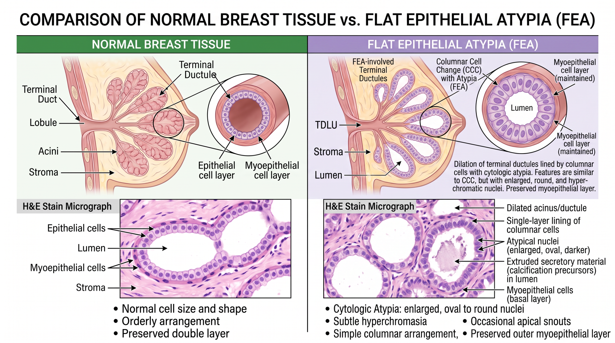
Flat epithelial atypia is a microscopic change in the small milk ducts and lobules of the breast. The lining cells become flatter or slightly enlarged, and their nuclei look a little more uniform and abnormal than usual. This is why a report may describe atypical breast cells, even though the finding is not invasive cancer and not ductal carcinoma in situ. A helpful analogy is a smoke detector that goes off before anyone sees a fire; the alarm deserves attention, but it does not mean a fire has already been found.
In the laboratory, the diagnosis depends on how the cells look on stained tissue slides from a core needle biopsy. Pathologists examine the architecture of the ducts, the shape of the cells, the nuclear features, and whether the biopsy also shows calcifications. Flat epithelial atypia is closely related to a spectrum of breast changes called columnar cell change, in which duct lining cells become taller or altered. When the cells also show mild atypia, the report may use the term flat epithelial atypia.
This breast biopsy result matters because a needle biopsy samples only part of the area seen on imaging. If the sampled tissue explains the imaging finding completely, the risk may be lower. If the imaging abnormality is larger, more suspicious, or not fully represented in the tissue, additional sampling may be recommended. Patients who want help understanding the structure and wording of the report may find Understanding Your Pathology Report: How to Read It with Confidence useful while preparing for the next appointment.
Why Your Report Shows This Finding
Flat epithelial atypia is often discovered because a screening mammogram shows tiny calcium deposits. These deposits are commonly called calcifications mammogram findings, and they can form in benign breast tissue, atypical areas, or cancerous areas. The biopsy is performed to learn what tissue change is causing the imaging appearance. When the microscope shows flat epithelial atypia and the calcifications are present in the sampled tissue, the pathology and radiology findings may be considered concordant.
A core needle biopsy removes several narrow pieces of tissue from the area of concern. Each piece is processed, embedded in paraffin, cut into very thin sections, stained, and reviewed under a microscope. The pathologist looks for columnar cell change, atypical breast cells, ductal carcinoma in situ, invasive carcinoma, and other benign breast conditions. The diagnosis is based on what is actually present in the tissue submitted to the laboratory, not on the mammogram alone.
Sometimes the report shows only flat epithelial atypia, and sometimes it appears with other findings, such as atypical ductal hyperplasia, lobular neoplasia, papilloma, radial scar, or usual ductal hyperplasia. These associated findings can change the management plan because they affect the chance that a more serious lesion may be nearby. The term upgrade rate refers to the percentage of cases that show ductal carcinoma in situ or invasive cancer when more tissue is removed after the needle biopsy. The upgrade rate is not the same for every patient, because it depends on imaging features, sampling amount, calcifications mammogram extent, and whether other atypical lesions are present.
How Serious Is Flat Epithelial Atypia?
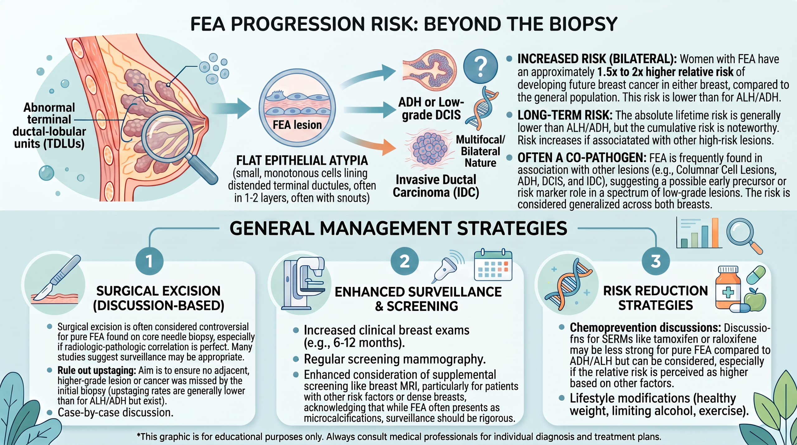
Flat epithelial atypia is serious enough to require a thoughtful follow-up plan, but it should not be understood as a cancer diagnosis. In many cases, it behaves as a marker that the breast tissue in that area has shown early atypical change. The key question is whether the core needle biopsy sampled the entire area of concern well enough. When the pathology finding matches the imaging finding and most calcifications were removed, the risk of finding cancer later may be low.
The seriousness of flat epithelial atypia increases when there is discordance between the imaging and the pathology. Discordance means the biopsy result does not adequately explain what the radiologist saw. For example, if calcifications mammogram findings looked highly suspicious but only a tiny amount of calcium was found in the biopsy tissue, the team may worry that the most important area was missed. In that setting, surgical excision or repeat biopsy may be recommended to obtain a fuller answer.
The presence of additional atypical breast cells can also change the level of concern. If atypical ductal hyperplasia, ductal carcinoma in situ, or another higher-risk lesion is present, management is different from isolated flat epithelial atypia. The upgrade rate is often discussed in this context because it helps estimate how often a larger tissue sample finds something more significant. A patient should not have to interpret that number alone; the most meaningful risk estimate comes from combining the breast biopsy result, imaging pattern, tissue sampling, and individual risk factors.
What Happens Next: Treatment and Monitoring
The next step after a diagnosis of flat epithelial atypia usually involves radiology-pathology correlation. This means the radiologist and pathology team compare the mammogram target with what was found in the core needle biopsy. If the biopsy contains the calcifications and the microscopic finding explains the imaging abnormality, some centers may recommend close imaging follow-up instead of surgery. If the sampling is limited or the imaging remains concerning, surgical excision may be recommended.
Surgical excision is not the same as breast cancer surgery. It is usually a diagnostic procedure designed to remove the area around the biopsy site so that the tissue can be examined more completely. The excision specimen allows the pathologist to look for any nearby ductal carcinoma in situ or invasive carcinoma that might not have been captured in the needle sample. When surgical excision shows only flat epithelial atypia or benign changes, the result can provide meaningful reassurance.
If monitoring is chosen, follow-up often includes a diagnostic mammogram at a defined interval, commonly around six months, followed by additional imaging based on institutional practice and risk assessment. The plan may also include a breast specialist visit to review personal risk factors, family history, breast density, and prior biopsies. This does not mean the patient has cancer; it means the care team is making sure the area remains stable. For general background on what a pathology report is and how tissue diagnoses are created, What Is a Pathology Report? offers a plain-language overview.
Questions to Ask Your Doctor or Pathologist
- Did the core needle biopsy remove the calcifications or imaging target that led to the biopsy?
- Is the breast biopsy result considered concordant with the mammogram or ultrasound findings?
- Was flat epithelial atypia the only abnormal finding, or were other atypical breast cells present?
- What is the estimated upgrade rate for this specific case based on imaging and sampling?
- Is surgical excision recommended, or is imaging follow-up a reasonable option?
- Were the calcifications identified in the tissue sections reviewed by the pathologist?
- Would a second pathology opinion change management before surgery or monitoring?
These questions are useful because flat epithelial atypia is a diagnosis that depends heavily on context. The same phrase can lead to different recommendations depending on sampling adequacy, calcifications mammogram extent, and associated pathology findings. A brief but focused conversation can often clarify why one plan is preferred over another.
A second review may be helpful when the report includes several high-risk lesions, when the imaging and pathology do not seem to match, or when surgical excision is being considered. Patients considering review can read When Should You Get a Second Pathology Opinion? or How to Get a Second Opinion on Your Pathology Diagnosis before requesting slides and records.
Frequently Asked Questions
Is flat epithelial atypia breast cancer?
No. flat epithelial atypia is not breast cancer, and it is not the same as invasive carcinoma. It is a high-risk or atypical microscopic finding that may sometimes be found near more significant disease. The main reason it receives attention is the possibility that a core needle biopsy sampled only part of the imaging abnormality.
Does flat epithelial atypia need surgery?
Not every case requires surgery. Some patients are offered surgical excision, especially when sampling is limited, imaging is concerning, or other atypical breast cells are present. Other patients may be monitored with imaging if the breast biopsy result is concordant and the target was well sampled. The decision depends on the full radiology-pathology picture.
What is the upgrade rate for flat epithelial atypia?
The upgrade rate is the chance that surgical excision finds ductal carcinoma in situ or invasive cancer after flat epithelial atypia was diagnosed on needle biopsy. Published estimates vary because studies include different imaging patterns, biopsy methods, and associated lesions. The most relevant upgrade rate is the one applied to the patient’s exact scenario. A breast specialist or pathologist can explain how local data and individual features affect that estimate.
Can flat epithelial atypia go away?
Flat epithelial atypia is a tissue diagnosis rather than a symptom that can be watched to “go away” in the usual sense. If the abnormal area was removed by the biopsy or by surgical excision, there may be no remaining lesion in that spot. If monitoring is chosen, imaging is used to make sure the area remains stable. Stability over time is reassuring, but follow-up should match the care team’s recommendation.
Should I get a second opinion for flat epithelial atypia?
A second opinion can be helpful if the recommendation is unclear, if surgery is planned, or if the pathology report includes multiple atypical findings. Breast pathology can involve subtle distinctions, especially between flat epithelial atypia, atypical ductal hyperplasia, and low-grade ductal carcinoma in situ. A review may confirm the diagnosis or refine the level of concern. It is reasonable to request review before making a major decision.
Flat epithelial atypia can sound frightening, but the diagnosis often represents a careful warning sign rather than cancer. The safest plan comes from matching the microscope findings with the imaging target and the amount of tissue sampled. Honest Pathology consultations can help patients and families understand the wording, the uncertainty, and the right questions to bring back to the treating team.
References:
National Cancer Institute — Pathology Reports Fact Sheet
National Cancer Institute — Biopsy
NCBI Bookshelf — Biomedical Literature and Clinical References
