
Worried by atypical ductal hyperplasia on a breast biopsy report? Learn what it means, why it matters, and what to ask next.
atypical ductal hyperplasia means that some breast duct cells look abnormal and are growing more than expected, but the biopsy does not show invasive breast cancer. This finding is usually described on a breast biopsy report after a core needle biopsy done for calcifications, a lump, or another mammogram finding. Pathologists classify it as a high risk lesion because it can be associated with nearby ductal carcinoma in situ or a higher future breast cancer risk.
Seeing the word “atypical” can feel like a warning siren, especially when the report arrives before a full explanation. The phrase often creates a painful gray zone: Not cancer, but not completely normal either. A clear explanation can make that gray zone less frightening and help patients prepare for a calm, focused conversation with the breast care team.
Pathology language is precise because treatment decisions depend on tiny differences under the microscope. A helpful starting point is understanding that atypical ductal hyperplasia is a diagnosis based on cell appearance, growth pattern, and the limited amount of tissue present in the biopsy sample.
Atypical Ductal Hyperplasia — What It Actually Means
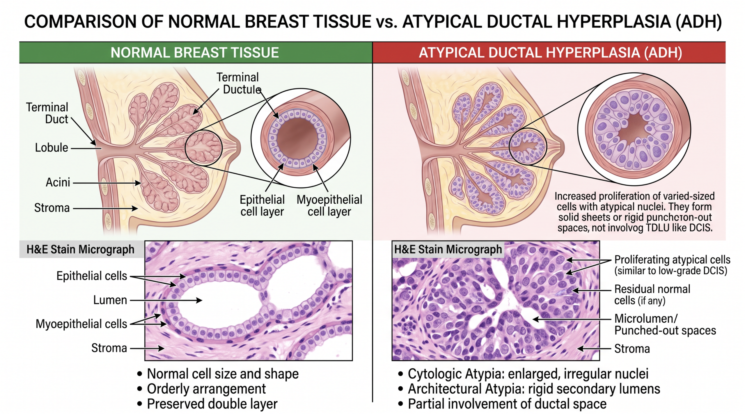
Atypical ductal hyperplasia is an overgrowth of abnormal-looking ductal cells inside the milk ducts of the breast. “Ductal” means the change begins in the ducts, the small channels that normally carry milk. “Hyperplasia” means extra cell growth. “Atypical” means the cells do not look completely normal when examined by a pathologist under the microscope.
A useful analogy is a smoke detector that goes off before a fire is visible. The alarm does not prove there is a fire, but it deserves attention because something unusual has been detected. In the same way, atypical ductal hyperplasia is not invasive breast cancer, but it is not ignored. It is called a high risk lesion because it can sit near a more advanced abnormality or signal increased breast cancer risk over time.
On a breast biopsy report, the diagnosis may appear as “ADH,” “atypical ductal hyperplasia,” or “focal atypical ductal hyperplasia.” The pathologist looks at whether the abnormal ductal cells have some features of low-grade ductal carcinoma in situ, often called DCIS, but not enough in size or extent to call it DCIS. This distinction can be subtle, especially in a core needle biopsy where only small tissue fragments are available. For patients trying to understand report wording, a structured approach to reading a pathology report can make the terminology feel less overwhelming.
Why Your Report Shows This Finding
Atypical ductal hyperplasia is most often found after a breast biopsy done for calcifications seen on mammography. These calcifications are tiny calcium deposits that can form in benign breast tissue, high risk lesion areas, DCIS, or invasive cancer. The mammogram finding helps the radiologist choose the area to sample, but the final diagnosis depends on microscopic review. A core needle biopsy removes small cylinders of tissue so the pathologist can evaluate the architecture and the ductal cells.
Under the microscope, ductal cells in atypical ductal hyperplasia tend to grow in a more uniform and organized way than usual hyperplasia. They may form small bridges, arcs, or rigid spaces inside ducts. These patterns overlap with low-grade DCIS, which is why the diagnosis can feel confusing. The difference may come down to how much of the duct is involved, how large the focus is, and whether the pattern is fully developed.
A breast biopsy report may also mention related findings such as columnar cell change, flat epithelial atypia, usual ductal hyperplasia, papilloma, radial scar, or calcifications. These findings can influence whether surgical excision is recommended. The report may also state whether the calcifications seen on imaging were present in the tissue sample, which helps confirm that the mammogram finding was sampled correctly. When the imaging result and pathology result do not seem to match, the care team may recommend additional sampling or review.
How Serious Is Atypical Ductal Hyperplasia?
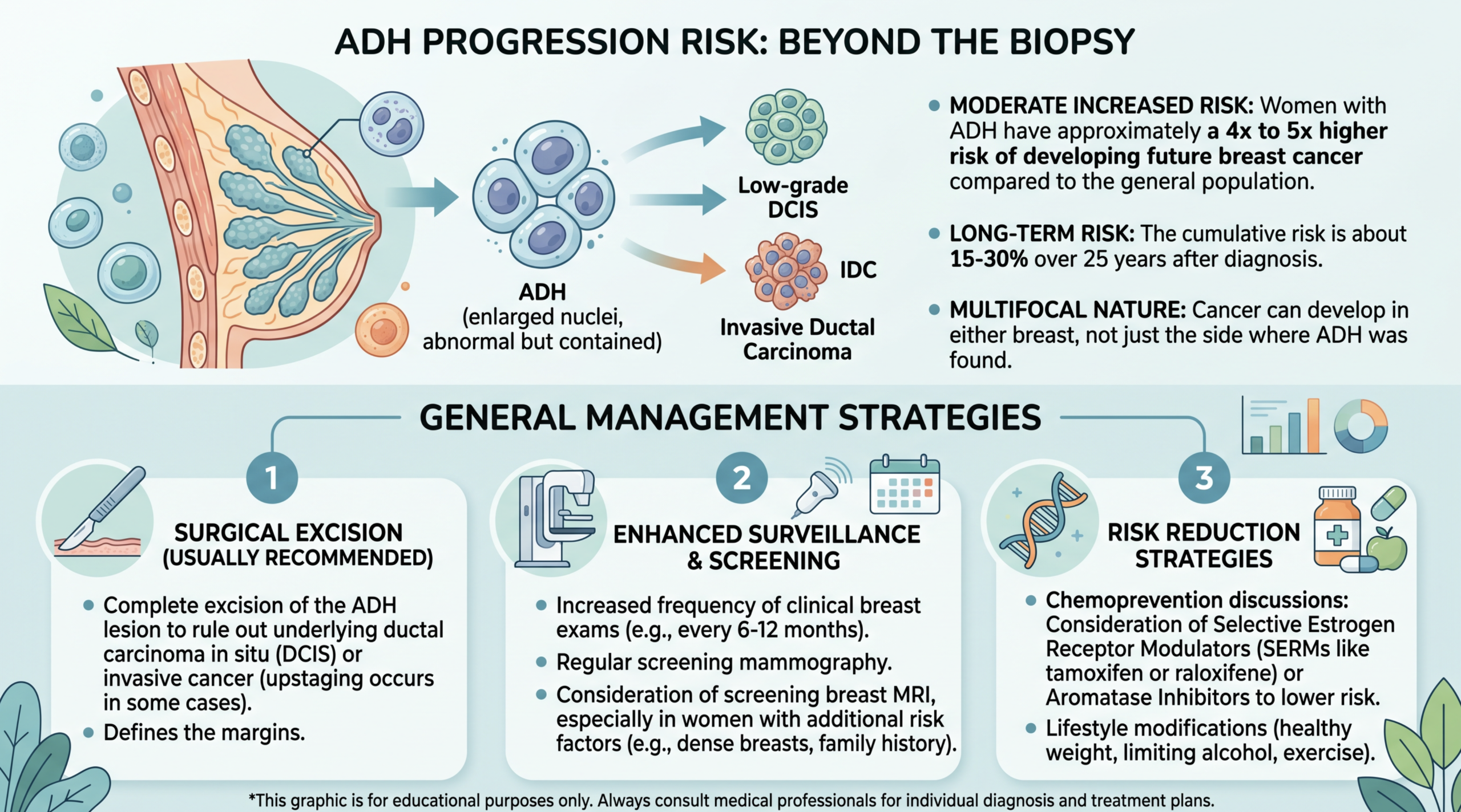
Atypical ductal hyperplasia is serious enough to require follow-up, but it is not the same as a diagnosis of invasive breast cancer. The seriousness comes from two related issues. First, a core needle biopsy samples only part of the abnormal area, so surgical excision may find DCIS or, less commonly, invasive cancer nearby. Second, atypical ductal hyperplasia is linked to higher breast cancer risk in either breast over time.
The risk is not the same for every patient. A tiny focus of atypical ductal hyperplasia that fully explains a small group of calcifications may be handled differently from a larger or more extensive abnormality. Radiology-pathology concordance matters, meaning the imaging findings and the breast biopsy report appear to fit together. If the mammogram finding looked more suspicious than the biopsy result explains, surgical excision becomes more important.
Many patients are told that atypical ductal hyperplasia is a high risk lesion rather than cancer. That wording is accurate, but it can still be emotionally difficult. In practical terms, a high risk lesion means the medical team wants to rule out a nearby higher-grade process and plan long-term surveillance. Breast cancer risk may also be estimated using personal history, family history, genetic factors, breast density, and the number of atypical areas found.
The most concerning scenario is when atypical ductal hyperplasia is extensive, present at multiple sites, associated with a suspicious mammogram finding, or found along with other atypical lesions. The more reassuring scenario is a small, well-sampled focus with imaging and pathology that match closely. Even in reassuring cases, careful follow-up is still typical. A second pathology opinion may be considered when the diagnosis is borderline between atypical ductal hyperplasia and DCIS, or when the treatment plan depends heavily on that distinction.
What Happens Next: Treatment and Monitoring
After atypical ductal hyperplasia is found, the next step is usually discussion with a breast surgeon or breast specialist. Many patients are advised to have surgical excision, also called an excisional biopsy or lumpectomy, to remove the remaining abnormal area. The purpose of surgical excision is not because the core diagnosis is cancer. The purpose is to make sure there is not a nearby DCIS or invasive cancer that the core needle biopsy did not capture.
If surgical excision shows only atypical ductal hyperplasia and no cancer, treatment often shifts toward risk management. This may include regular mammograms, sometimes breast MRI for selected higher-risk patients, and clinical breast exams. Some patients may be referred to a high-risk breast clinic. Discussions may include lifestyle factors, family history, genetic counseling, and medications that can reduce breast cancer risk in carefully selected patients.
Monitoring is individualized. A patient with atypical ductal hyperplasia, strong family history, and dense breasts may have a different plan than a patient with a small isolated focus and no additional risk factors. The breast biopsy report is one piece of the larger picture, along with imaging and clinical history. For patients new to pathology terminology, understanding what a pathology report is can help explain why microscopic findings carry so much weight in care decisions.
Questions to Ask Your Doctor or Pathologist
- Does the breast biopsy report show atypical ductal hyperplasia only, or are there other high risk lesion findings?
- Were the calcifications or the mammogram finding successfully sampled in the core needle biopsy?
- Is the imaging result concordant with the pathology result?
- Is surgical excision recommended, and what is the reason in this specific case?
- What is the chance that surgical excision could find DCIS or invasive cancer?
- How does this diagnosis affect long-term breast cancer risk?
- Should the slides be reviewed by a breast pathologist before surgery or treatment decisions?
These questions can help turn a frightening report into a practical conversation. Atypical ductal hyperplasia often requires teamwork among radiology, pathology, surgery, and breast oncology, and each specialty contributes a different part of the answer.
A second opinion can be especially helpful when the wording is borderline, when the biopsy is small, or when surgical excision is being planned. Patients considering review may find guidance on when to get a second pathology opinion useful before the next appointment.
Frequently Asked Questions
Is atypical ductal hyperplasia breast cancer?
No. Atypical ductal hyperplasia is not invasive breast cancer, and it is not automatically DCIS. It is a high risk lesion made up of abnormal ductal cells growing inside breast ducts. The concern is that a core needle biopsy samples only part of the area. For that reason, surgical excision may be recommended to check for a more advanced nearby finding.
Can atypical ductal hyperplasia go away?
Atypical ductal hyperplasia is a tissue diagnosis, not a temporary blood test result. The area sampled by the biopsy does not simply “normalize” in a way that can be confirmed without tissue evaluation. If the area is removed by surgical excision and no cancer is found, the immediate abnormal focus may be gone. However, breast cancer risk can remain higher than average over time.
Why do I need surgery if atypical ductal hyperplasia is not cancer?
Surgical excision is often recommended because the core needle biopsy may not show the entire abnormal area. Sometimes surgical excision finds only atypical ductal hyperplasia, which is reassuring. In other cases, it may find DCIS or a small invasive cancer nearby. The decision depends on the breast biopsy report, imaging appearance, and whether the mammogram finding was fully explained.
How often does atypical ductal hyperplasia become cancer?
Atypical ductal hyperplasia does not always “turn into” cancer in a straight-line way. Instead, it marks breast tissue that has a higher breast cancer risk than average. Some studies show an increased long-term risk, and some excision specimens show DCIS or invasive cancer already present nearby. The exact risk depends on the amount of atypia, imaging findings, age, family history, and other clinical factors.
Should I get a second opinion for atypical ductal hyperplasia?
A second opinion can be reasonable because atypical ductal hyperplasia can overlap microscopically with low-grade DCIS. Review by a breast pathologist may be especially helpful if the diagnosis is borderline, if treatment recommendations differ, or if surgery is being planned. A second opinion may confirm the diagnosis, refine the wording, or identify a more specific category. Practical steps are outlined in how to get a second opinion on a pathology diagnosis.
Atypical ductal hyperplasia can feel frightening because it lives between normal tissue and cancer terminology. The most helpful path forward is clear confirmation of the diagnosis, careful matching with imaging, and a plan tailored to the patient’s personal risk. Honest Pathology consultations can support patients and families who want an expert review of the pathology wording before major decisions are made.
References:
National Cancer Institute — Pathology Reports Fact Sheet
National Cancer Institute — Biopsy Definition
NCBI Bookshelf — Medical Reference Library
