
Worried after a radial scar breast biopsy? Learn what it means, why excision may be advised, and what to ask the care team next.
Radial scar breast biopsy usually means a noncancerous breast finding, but it often needs careful review because it can mimic cancer on imaging and may occur near higher-risk changes.
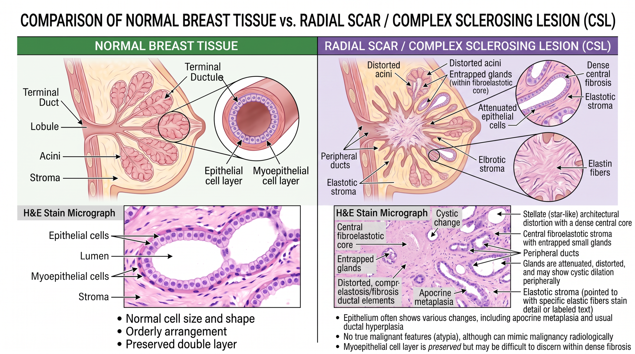
Pathologists use this diagnosis when breast ducts and glands are pulled into a scar-like center, creating a star-shaped pattern under the microscope. The word “scar” can sound reassuring, while the words “complex sclerosing lesion” can sound frightening, and both terms often refer to the same general process.
Receiving this result can feel confusing because the report may say “benign” but the radiologist or surgeon may still discuss additional sampling or surgery. That apparent contradiction is one of the hardest parts for patients and families. The goal is not to create alarm; it is to make sure the small tissue sample truly represents the entire imaging abnormality.
Radial Scar Breast Biopsy — What It Actually Means
A radial scar breast biopsy diagnosis describes a benign, scar-like breast lesion with ducts and glands arranged around a central fibrotic area. Despite the name, it usually is not a scar from an injury, surgery, or trauma. It is a microscopic pattern in breast tissue that can look spiky or star-shaped, especially when it causes architectural distortion on mammography. A complex sclerosing lesion is generally a larger or more elaborate version of the same type of finding.
A helpful analogy is a wrinkle in fabric. From the outside, the wrinkle may create shadows and pulled lines that resemble a tear, even when the fabric itself is not torn. In the breast, a radial scar can pull nearby tissue inward, creating architectural distortion that resembles invasive cancer on imaging. That is why a benign breast biopsy report can still lead to a recommendation for more evaluation.
Under the microscope, the pathologist checks whether the glands still have the normal two-cell layer that supports a benign interpretation. Immunohistochemistry may be used when the pattern is difficult to separate from invasive carcinoma. The report may also mention proliferative changes, usual ductal hyperplasia, papilloma, or atypical hyperplasia. For a patient reading a breast biopsy report, the key question is whether the radial scar breast biopsy finding is present alone or with atypia or another high risk lesion.
Why the Report Shows This Finding
A radial scar or complex sclerosing lesion is often found because breast imaging shows architectural distortion, calcifications, or a mass-like area. Sometimes it is too small to feel on physical examination. A core needle biopsy is then performed to sample the area that looks abnormal on imaging. The pathology diagnosis is based on the tissue cores received in the laboratory, not on imaging alone.
In the lab, the pathologist examines the core needle biopsy tissue in thin sections on glass slides. The central scar-like zone, trapped ducts, and radiating gland pattern support the diagnosis. The pathologist also searches carefully for atypical hyperplasia, ductal carcinoma in situ, lobular neoplasia, or invasive cancer. These additional findings matter because they can change the meaning of the breast biopsy report and the next step in care.
The reason this diagnosis receives extra attention is sampling. A core needle biopsy removes only narrow pieces of tissue from a larger imaging target. If the sampled tissue shows a complex sclerosing lesion but imaging looks more worrisome, the care team may be concerned that a nearby abnormal area was missed. This is called radiology-pathology correlation, and it means the imaging findings and pathology findings must make sense together.
How Serious Is a Radial Scar Breast Biopsy?
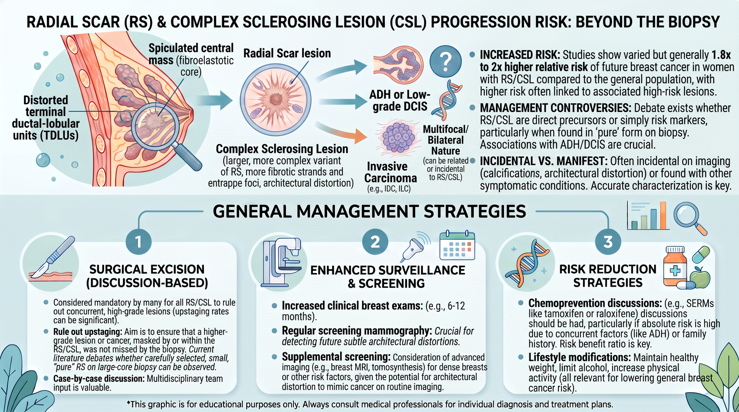
A radial scar breast biopsy is serious enough to deserve careful follow-up, but it is not the same as being told that breast cancer is present. When the radial scar is small, completely explained by imaging, and has no atypical hyperplasia, many cases are managed with either close imaging follow-up or surgical excision, depending on local practice and patient factors. The highest concern occurs when the lesion is associated with atypia, discordant imaging, or incomplete sampling. In that setting, the chance of finding a more significant lesion nearby is higher.
The word “upgrade” is often used in this context. An upgrade means that surgical excision or additional sampling finds ductal carcinoma in situ, invasive carcinoma, or a higher-risk diagnosis that was not present in the original core needle biopsy. Upgrade risk varies across studies because lesions differ in size, imaging appearance, biopsy method, and whether atypia is present. A radial scar breast biopsy without atypia usually carries a lower upgrade risk than a radial scar with atypical hyperplasia.
For many patients, the most useful way to think about this diagnosis is as a marker that the area needs confirmation, not as a cancer diagnosis by itself. A high risk lesion does not mean cancer is already there; it means the tissue pattern is linked to a higher chance of nearby or future abnormality compared with completely ordinary breast tissue. A complex sclerosing lesion can sit in this gray zone, where the pathology is benign but the decision-making remains cautious. When there is uncertainty, a second pathology review can help confirm whether the sampled tissue truly shows only a radial scar breast biopsy finding or whether another diagnosis is present.
What Happens Next: Treatment and Monitoring
After a radial scar breast biopsy, the next step usually depends on three things: the pathology details, the imaging appearance, and whether the findings are concordant. Concordant means the biopsy result adequately explains what the radiologist saw. If the imaging target was architectural distortion and the biopsy shows a radial scar or complex sclerosing lesion, the result may be considered concordant. If the imaging looked highly suspicious but the tissue sample shows only minimal benign change, more sampling may be recommended.
Surgical excision is commonly discussed because it removes the entire targeted area and allows the pathologist to examine more tissue. In some centers, small radial scars without atypical hyperplasia and with excellent sampling may be followed with imaging instead. Other centers recommend surgical excision more routinely, especially for larger lesions, limited sampling, or a high risk lesion. The choice should be individualized and based on the complete breast biopsy report, imaging report, and clinical history.
If surgery is performed, the excision specimen is processed carefully to look for any hidden ductal carcinoma in situ or invasive cancer. If no cancer or atypia is found, the result is reassuring, and follow-up usually returns to breast imaging surveillance. If atypical hyperplasia or another high risk lesion is found, risk assessment and prevention discussions may follow. If cancer is found, treatment planning shifts to the standard breast cancer pathway, with decisions guided by the exact diagnosis, size, margins, receptors, and stage.
Questions to Ask the Doctor or Pathologist
- Was the radial scar breast biopsy result concordant with the mammogram, ultrasound, or MRI finding?
- Does the breast biopsy report mention atypical hyperplasia, ductal carcinoma in situ, lobular neoplasia, or invasive cancer?
- Was the lesion called a radial scar, a complex sclerosing lesion, or both?
- How large was the imaging abnormality, and how much tissue was sampled by core needle biopsy?
- Is surgical excision recommended, or is imaging follow-up considered safe in this specific case?
- What is the estimated upgrade risk based on the exact pathology and imaging findings?
- Would a second pathology opinion change management before surgery or surveillance?
These questions help turn a frightening phrase into a practical plan. They also make clear that the diagnosis is not interpreted in isolation; radiology, pathology, and clinical history all matter.
A second review can be especially helpful when the report mentions atypia, when imaging and pathology do not match, or when surgical excision is being weighed against surveillance. Helpful background about when review may be appropriate is available in When Should You Get a Second Pathology Opinion? and How to Get a Second Opinion on Your Pathology Diagnosis.
Frequently Asked Questions
Is a radial scar in the breast cancer?
A radial scar in the breast is usually not cancer. A radial scar breast biopsy most often describes a benign scar-like pattern in breast tissue. The concern is that it can look like cancer on imaging and can sometimes occur near atypical hyperplasia, ductal carcinoma in situ, or invasive cancer. That is why follow-up, correlation, or surgical excision may be recommended even when the biopsy itself is benign.
Why does a benign radial scar need surgery?
Surgical excision may be recommended because a core needle biopsy samples only part of the imaging abnormality. If the entire area is removed, the pathologist can examine more tissue and exclude a nearby cancer or high risk lesion. This recommendation does not mean cancer has already been found. It means the care team wants a more complete answer before choosing long-term surveillance.
What is the difference between radial scar and complex sclerosing lesion?
A complex sclerosing lesion is closely related to a radial scar and is often a larger or more complex version of the same process. Both can create architectural distortion and both can appear suspicious on breast imaging. The breast biopsy report may use one term or both terms depending on the size and microscopic appearance. Management depends less on the name alone and more on atypia, imaging concordance, and sampling.
Can a radial scar breast biopsy be wrong?
A radial scar breast biopsy can be limited by sampling, because the needle removes only selected tissue cores. The pathologist may correctly diagnose a radial scar in the sampled tissue, while a nearby unsampled area could contain atypical hyperplasia or cancer. This is why radiology-pathology correlation matters so much. If the imaging and pathology do not match well, additional biopsy, surgical excision, or second pathology review may be advised.
Does radial scar increase breast cancer risk?
A radial scar may be associated with a slightly increased breast cancer risk, especially when it occurs with atypical hyperplasia or other proliferative changes. The risk is not the same for every patient and should be interpreted with age, family history, imaging findings, and the complete pathology report. A high risk lesion found with the radial scar may lead to more formal breast risk assessment. The care team may recommend enhanced screening or prevention counseling depending on the full picture.
A radial scar or complex sclerosing lesion can be unsettling because the words sound serious and the plan may not be simple. Clear correlation between imaging and pathology is the safest way forward. Honest Pathology consultations can provide an independent review of the slides and report when patients or clinicians need added clarity before excision, surveillance, or a second opinion decision.
References:
National Cancer Institute — Pathology Reports
National Cancer Institute — Biopsy
NCBI Bookshelf — Medical Reference Texts
