
A breast biopsy can feel frightening when atypical lobular hyperplasia appears with LCIS. Learn the difference and what to ask next.
Atypical lobular hyperplasia is a breast biopsy finding that means some cells in the milk-producing lobules look abnormal, but the finding is not the same as invasive breast cancer.
It belongs to a group of changes called lobular neoplasia, which also includes lobular carcinoma in situ. These diagnoses can sound alarming because the words “atypical” and “carcinoma” appear in the report, yet their meaning depends heavily on the amount of abnormal change seen under the microscope.
Receiving this result can make a patient feel as if a trapdoor opened beneath an ordinary screening mammogram or biopsy. Clear pathology language matters because the next step may be observation, surgical excision, imaging follow-up, risk-reduction medication, or a second pathology review, depending on the exact report and clinical context.
Atypical Lobular Hyperplasia — What It Actually Means
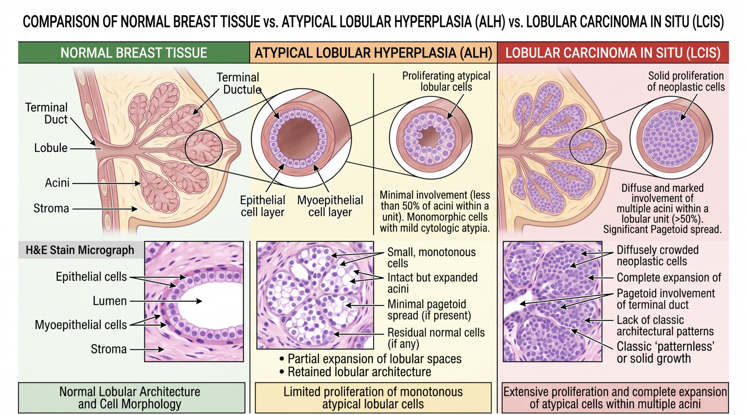
Atypical lobular hyperplasia describes a small, limited overgrowth of abnormal-looking cells inside breast lobules. Lobules are the tiny glands that can make milk, and they are lined by cells that usually form orderly layers. In atypical lobular hyperplasia, some of those cells become loosely attached, mildly enlarged, and more crowded than expected. Pathologists often recognize this pattern because the cells fill part of a lobule but do not expand it enough to meet criteria for lobular carcinoma in situ.
A helpful analogy is a closet that has become partly overfilled. In atypical lobular hyperplasia, the closet has extra items and looks disorganized, but it is not completely packed from wall to wall. In lobular carcinoma in situ, the closet is more fully expanded by similar abnormal cells. This is why a breast biopsy report may describe both findings in the same family of diagnoses, while still separating them by extent.
The difference is microscopic, not based on how a patient feels. Atypical lobular hyperplasia usually does not cause a lump, pain, nipple change, or visible breast change by itself. It is often found on a core needle biopsy performed for calcifications, an imaging abnormality, or another nearby lesion. For patients trying to understand the wording, a general overview of what a pathology report contains can make the terminology less intimidating.
Why a Breast Biopsy Report Shows This Finding
A breast biopsy report shows atypical lobular hyperplasia when the sampled tissue contains small lobules filled by discohesive, abnormal epithelial cells. “Discohesive” means the cells do not stick together tightly, which is a classic feature of lobular neoplasia. Pathologists examine the architecture, the amount of lobule involvement, and whether the abnormal cells remain confined within the lobules. E-cadherin staining may be used when the pattern is subtle or when ductal and lobular changes need to be separated.
E-cadherin staining is an immunohistochemistry test that helps show whether breast cells are behaving more like ductal cells or lobular cells. Lobular lesions often show loss or marked reduction of E-cadherin staining, supporting a diagnosis within the lobular neoplasia spectrum. This does not mean the stain alone makes the diagnosis. It is interpreted together with the H&E slide appearance, imaging findings, and the type of sample, especially when the specimen comes from a core needle biopsy.
Atypical lobular hyperplasia and lobular carcinoma in situ may be discovered near benign changes such as fibrocystic change, columnar cell change, fibroadenoma, or calcifications. Sometimes they are incidental findings, meaning the biopsy was done for something else and the lobular change was found nearby. Other times, the imaging target and the pathology finding must be carefully matched to make sure the sampled tissue explains the mammogram or MRI abnormality. This radiology-pathology correlation is one reason a breast biopsy report is more than a list of words; it is part of a larger clinical puzzle.
How Serious Is Atypical Lobular Hyperplasia?
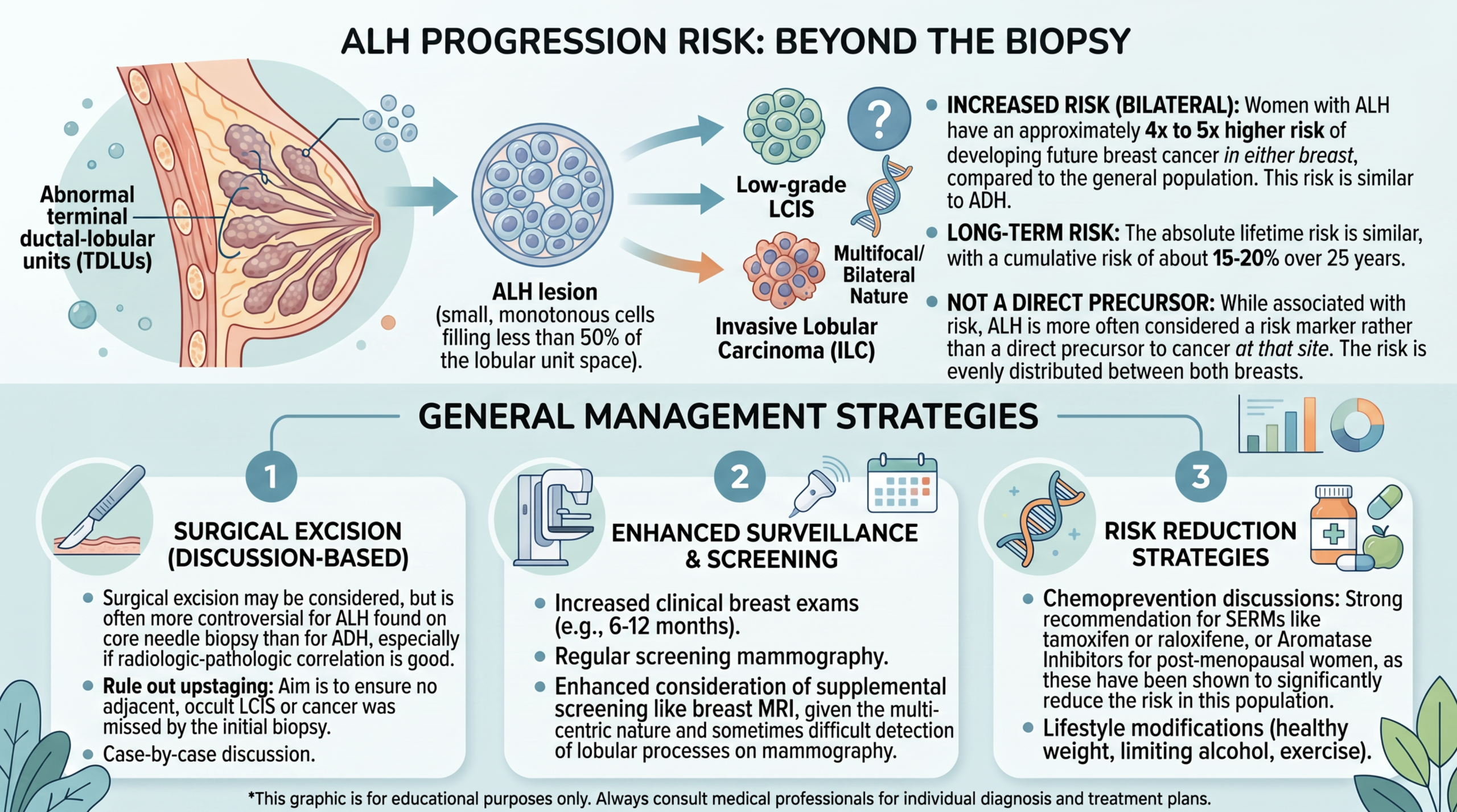
Atypical lobular hyperplasia is serious enough to deserve careful follow-up, but it is not the same as a diagnosis of invasive breast cancer. The main concern is that atypical lobular hyperplasia is a marker of increased future breast cancer risk in either breast. It also may occasionally sit near a more significant lesion that was not fully sampled by the original core needle biopsy. That is why the seriousness depends on the imaging target, biopsy method, amount of abnormal tissue, and whether the pathology result fully explains the imaging finding.
Compared with lobular carcinoma in situ, atypical lobular hyperplasia usually represents a smaller and more limited abnormal cell population. Classic lobular carcinoma in situ generally involves more lobules and expands them more completely, while pleomorphic LCIS is a more concerning variant that behaves differently and is managed more aggressively. A report that says classic LCIS is not the same as a report that says pleomorphic LCIS. This distinction is critical because breast cancer risk and surgical excision recommendations can change depending on the exact type.
In many cases, atypical lobular hyperplasia leads to a discussion about enhanced screening, risk assessment, and sometimes surgical excision. If imaging and pathology are concordant and the lesion is small, some teams may consider close imaging follow-up rather than immediate surgery. If there is discordance, a mass lesion, extensive involvement, another high-risk lesion, or uncertainty about sampling, surgical excision may be recommended. A second review can be especially helpful when the wording is borderline between atypical lobular hyperplasia, lobular carcinoma in situ, and another high-risk breast lesion.
ALH Versus LCIS: What Pathologists Look For
The distinction between atypical lobular hyperplasia and lobular carcinoma in situ is based mainly on how much of the lobular unit is filled and expanded by the abnormal cells. In classic teaching, atypical lobular hyperplasia involves part of a lobular unit, while lobular carcinoma in situ involves and expands more of it. The cells can look very similar, so the difference is often quantitative rather than dramatic. This is why two reports may use related terms, such as “lobular neoplasia,” when the border between categories is not perfectly sharp.
Pathologists also look for whether the lesion is classic or non-classic. Classic lobular carcinoma in situ has small, uniform cells that remain inside lobules and ducts, while pleomorphic LCIS has more enlarged, irregular cells and may show necrosis or calcifications. Pleomorphic LCIS is not managed like ordinary atypical lobular hyperplasia. When a breast biopsy report includes “pleomorphic,” “florid,” “necrosis,” or “mass-forming,” the clinical team usually pays closer attention to complete removal and imaging correlation.
Core needle biopsy sampling adds another layer of complexity. A core needle biopsy removes thin tissue cylinders, not the entire area seen on imaging. If atypical lobular hyperplasia is present only in the cores, the remaining breast tissue around the biopsy site is not fully visible to the pathologist. This is one reason surgical excision may be considered, particularly when the imaging abnormality is suspicious, the lesion is extensive, or the pathology does not fully account for the radiology finding.
What Happens Next: Treatment and Monitoring
The next step after atypical lobular hyperplasia usually begins with radiology-pathology correlation. The breast radiologist and treating clinician compare the imaging abnormality with the biopsy result to decide whether the finding is concordant. If the biopsy was performed for calcifications and the pathology explains those calcifications, follow-up may be different than if the biopsy was performed for a mass and only limited lobular neoplasia was found. This decision is individualized rather than automatic.
Surgical excision may be recommended to make sure no nearby ductal carcinoma in situ, invasive carcinoma, or more extensive lobular carcinoma in situ was missed. Surgical excision does not mean cancer has already been found. It means the team wants a larger tissue sample to rule out an upgrade. When surgical excision shows only atypical lobular hyperplasia or classic lobular carcinoma in situ, later care often focuses on breast cancer risk reduction and surveillance.
Monitoring may include annual mammography, possible breast MRI for higher-risk patients, formal risk calculation, lifestyle counseling, and discussion of medications that lower estrogen-driven breast cancer risk. A breast specialist may ask about family history, prior biopsies, breast density, genetic testing, and personal health factors. Patients reading a report line by line may benefit from learning how to read a pathology report with confidence. The goal is not to memorize every term, but to understand what the diagnosis does and does not mean.
Questions to Ask the Doctor or Pathologist
- Does the report say atypical lobular hyperplasia, classic lobular carcinoma in situ, pleomorphic LCIS, or another type of lobular neoplasia?
- Was the biopsy result concordant with the mammogram, ultrasound, or MRI finding?
- Was E-cadherin staining performed, and did it support a lobular diagnosis?
- Was the finding present in one core or in multiple cores from the core needle biopsy?
- Is surgical excision recommended because of sampling, imaging discordance, or another high-risk lesion?
- How does this diagnosis change breast cancer risk compared with the general population?
- Should the slides be reviewed by a breast pathologist before surgery or long-term surveillance is chosen?
These questions help turn a frightening breast biopsy report into a more organized discussion. A patient does not need to challenge the medical team to ask for clarification; good care includes making the diagnosis understandable. When atypical lobular hyperplasia and lobular carcinoma in situ are close possibilities, precise wording can affect both anxiety and management.
A second pathology opinion may be useful when the report uses borderline language, when the diagnosis was made on a small core needle biopsy, or when surgery is being considered. More information about when review is reasonable is available in this patient-focused article on when to get a second pathology opinion. If a review is pursued, the original glass slides, blocks if needed, imaging report, and final pathology report are usually the key materials.
Frequently Asked Questions
Is atypical lobular hyperplasia breast cancer?
Atypical lobular hyperplasia is not invasive breast cancer. It is a high-risk breast lesion and part of the lobular neoplasia spectrum. The finding means abnormal cells are present in lobules, but they have not invaded surrounding breast tissue. It does increase future breast cancer risk, so follow-up should be taken seriously.
What is the difference between ALH and LCIS?
The difference between ALH and LCIS is mainly the amount of lobular involvement seen under the microscope. Atypical lobular hyperplasia is more limited, while lobular carcinoma in situ fills and expands lobules more extensively. The cells may look very similar, which is why the distinction can be subtle. A breast pathologist may use E-cadherin staining and the overall slide pattern to support the diagnosis.
Does atypical lobular hyperplasia need surgery?
Atypical lobular hyperplasia does not always require surgery, but surgical excision is often discussed. The decision depends on imaging-pathology concordance, the amount of disease in the biopsy, the biopsy method, and whether other high-risk findings are present. If the core needle biopsy sample may not represent the whole imaging abnormality, excision may be recommended. If everything is concordant and limited, careful imaging follow-up may be considered by some teams.
Can LCIS turn into invasive cancer?
Classic lobular carcinoma in situ is best understood as both a risk marker and, in some cases, a possible non-obligate precursor. This means it increases breast cancer risk but does not guarantee that invasive cancer will develop. Pleomorphic LCIS is more concerning than classic LCIS and is usually managed more aggressively. The exact wording in the pathology report matters greatly.
Should I get a second opinion for atypical lobular hyperplasia?
A second opinion can be reasonable for atypical lobular hyperplasia, especially when the report is borderline with lobular carcinoma in situ or when surgery is being planned. Breast pathology distinctions can be nuanced, and another expert review may confirm the diagnosis or clarify the subtype. A patient can learn about the practical process through this article on how to get a second opinion on a pathology diagnosis. The goal is clearer decision-making, not creating delay or confusion.
Atypical lobular hyperplasia and LCIS can sound more frightening than they are when the report is read without context. The key is to separate “not invasive cancer” from “not important,” because this diagnosis deserves thoughtful risk assessment and follow-up. Honest Pathology consultations can help patients and families understand the exact wording of a report before major decisions are made.
References:
National Cancer Institute — Pathology Reports Fact Sheet
National Cancer Institute — Lobular Carcinoma in Situ Definition
National Cancer Institute — Biopsy Definition
