A scary report phrase can feel overwhelming. Learn what spindle cell neoplasm means, why classification is difficult, and what to ask next.
spindle cell neoplasm means that the pathologist sees a growth made of elongated cells, but the exact diagnosis may still need more testing and clinical context. This wording can appear in a pathology report after a biopsy or surgery, especially when a tumor has features that overlap with several different conditions. Some spindle cell tumors are benign, some are borderline, and some are malignant, so the phrase is a starting point rather than a final answer.
Seeing this term can be frightening because it sounds technical and unfinished. That fear is understandable. Pathology reports often use precise language to show what is known, what is suspected, and what still needs to be clarified. The goal is not to leave a patient in limbo; the goal is to avoid overcalling or undercalling a diagnosis before the evidence is complete.
Spindle cell tumors can be difficult because many unrelated diseases can look similar under the microscope. A careful review usually combines the microscope pattern, the biopsy sample location, immunohistochemistry stains, imaging findings, and sometimes molecular testing.
Spindle Cell Neoplasm — What It Actually Means
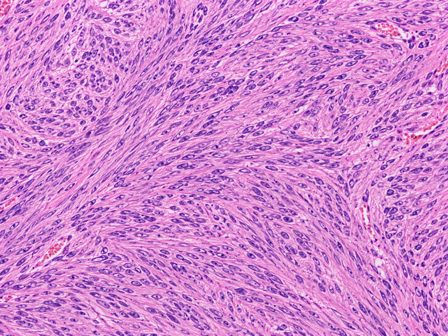
A spindle cell neoplasm is a tumor or growth made mostly of spindle cells, which are long, narrow cells that look somewhat like tiny stretched ovals under the microscope. The word “neoplasm” simply means a new growth. It does not automatically mean cancer. In a pathology report, this wording often tells the treating team that the pathologist has identified a real growth, but that the exact tumor type still needs to be classified.
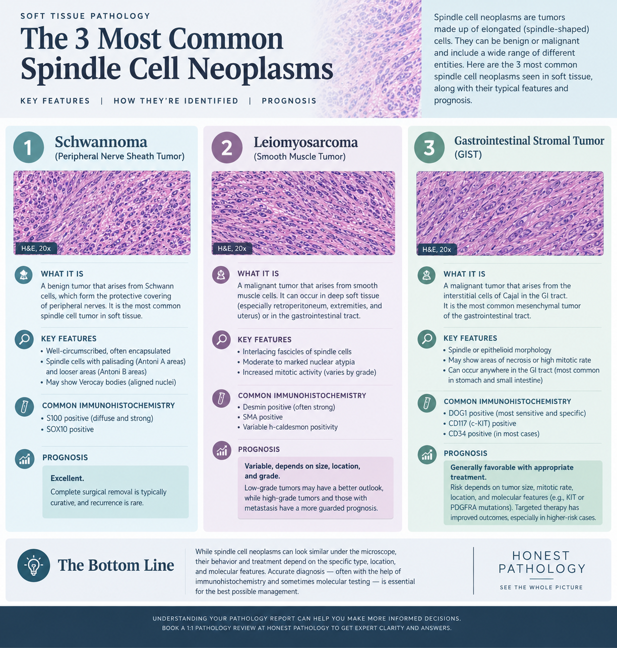
A helpful analogy is a car seen from far away through fog. The outline may show that it is definitely a vehicle, but the make, model, and condition may not be clear until the view improves. In the same way, spindle cells can form many different tumors, including benign nerve sheath tumors, fibrous tumors, smooth muscle tumors, inflammatory lesions, and sarcomas. The microscope gives important clues, but the final name may require more information.
This is why a pathology report may use terms such as “spindle cell lesion,” “spindle cell proliferation,” or spindle cell neoplasm. These phrases are not all identical, but they often reflect diagnostic caution. A soft tissue tumor in the arm, a similar-looking tumor in the uterus, and a similar-looking tumor in the skin may have very different meanings. Location, size, growth pattern, and patient history all matter.
Why Your Report Shows This Finding
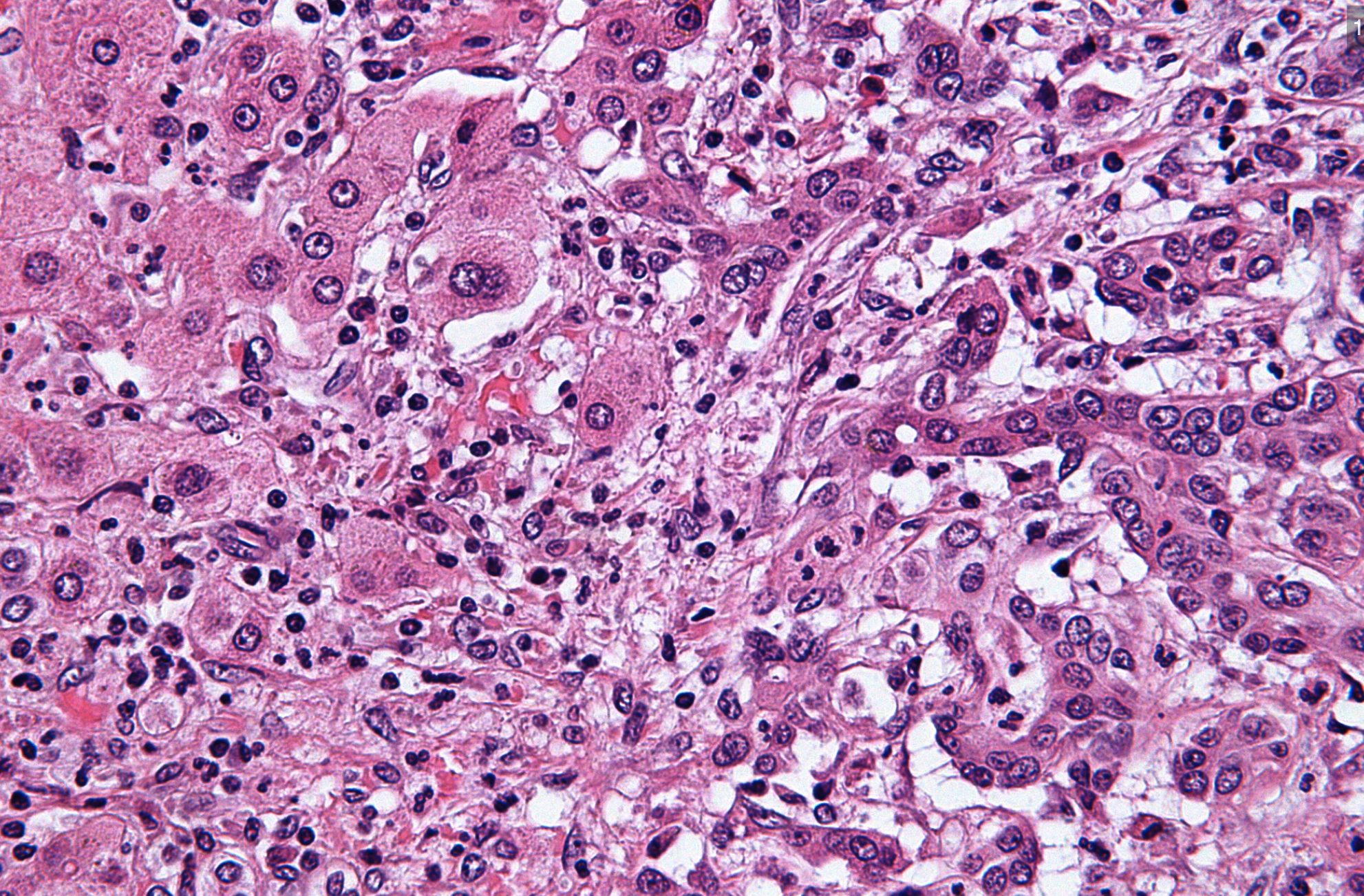
A report shows this finding when the pathologist sees a dominant population of spindle cells in the biopsy sample. These cells may form fascicles, sheets, whorls, or haphazard bundles. The pathologist looks at how uniform the cells are, how many mitoses are present, whether necrosis is seen, and whether the tumor invades nearby tissue. These features help separate a harmless process from a more concerning one.
Classification is difficult because spindle cells are a shared shape, not a single diagnosis. Smooth muscle tumors, nerve sheath tumors, fibrous tumors, gastrointestinal stromal tumors, melanoma, carcinoma with spindle features, and true sarcoma diagnosis categories can overlap. A soft tissue tumor may also look different depending on whether the biopsy sample is small, crushed, inflamed, or taken from only one part of a larger mass. A small biopsy sample may show the “architecture” of the tumor poorly, like judging an entire building from one brick.
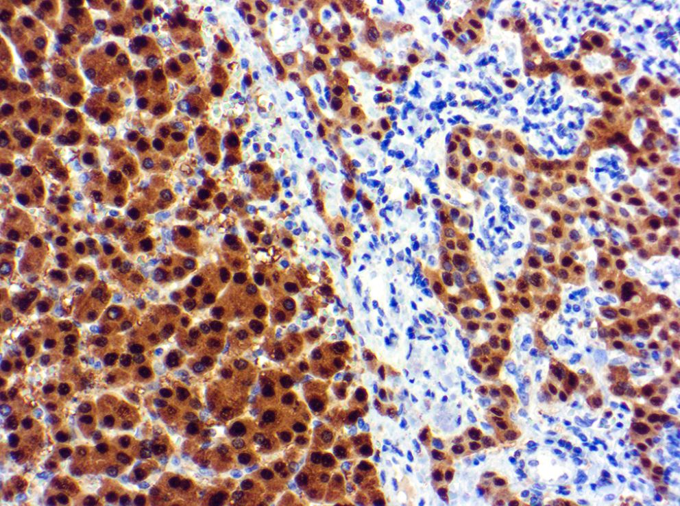
Immunohistochemistry stains are often ordered to narrow the possibilities. These tests use antibodies to look for proteins that point toward a line of differentiation, such as muscle, nerve, epithelial, vascular, or melanocytic origin. Molecular testing may be added when a tumor type is associated with a characteristic gene fusion, mutation, or chromosomal change. Immunohistochemistry stains and molecular testing do not replace the microscope impression; they add evidence to it.
How Serious Is Spindle Cell Neoplasm?
The seriousness of spindle cell neoplasm depends on the final diagnosis, not the phrase alone. Some are benign and removed completely with surgery. Others are locally aggressive, meaning they can come back in the same area even if they do not commonly spread. A smaller group are malignant tumors that require staging, specialized surgery, radiation, chemotherapy, targeted therapy, or a combination of treatments.
Pathologists often look for warning features when evaluating spindle cell neoplasm. These include marked atypia, a high mitotic rate, tumor necrosis, infiltrative edges, and certain immunohistochemistry stains that support a malignant diagnosis. In a possible sarcoma diagnosis, the tumor grade, size, depth, location, and margin status become very important. A pathology report may also mention Ki-67, which estimates how actively tumor cells are dividing; patients who see that term may find additional background in this explanation of Ki-67.
It is also possible for spindle cell neoplasm to be used before the pathologist is ready to commit to one category. That does not mean the situation is being ignored. It means the pathologist is protecting diagnostic accuracy by collecting more evidence. For many patients, the next report addendum or expert review provides the more specific name that guides treatment.
What Happens Next: Treatment and Monitoring
The next step depends on how much information is already available. If the biopsy sample is small, the treating physician may recommend complete excision, repeat biopsy, imaging, or referral to a specialist center. If the tumor is already removed, the pathology report may focus on margin status, tumor size, depth, grade, and whether the tumor has high-risk features. For a soft tissue tumor, imaging often helps determine whether the disease is confined to one site.
Additional testing commonly includes immunohistochemistry stains, molecular testing, or review by a pathologist with expertise in soft tissue pathology. A second review can be especially useful when the report uses broad terminology, when the diagnosis will change treatment, or when the tumor is rare. Patients who are trying to understand the structure of the report may benefit from a practical guide to reading pathology reports. For general background on what each section means, this overview of a pathology report may also help.
Treatment varies widely. A benign spindle cell tumor may need only surgery and follow-up. A malignant soft tissue tumor may require care from a multidisciplinary team, often including surgical oncology, medical oncology, radiation oncology, radiology, and pathology. Monitoring may include physical exams and imaging at intervals based on the final diagnosis and risk level.
Questions to Ask Your Doctor or Pathologist
- What is the most likely specific diagnosis for this spindle cell neoplasm?
- Was the biopsy sample large enough to classify the tumor confidently?
- Were immunohistochemistry stains performed, and what did they show?
- Is molecular testing recommended for this tumor type?
- Does the pathology report suggest a benign tumor, borderline tumor, or malignant tumor?
- If the tumor was removed, are the surgical margins clear?
- Would review by a soft tissue pathology specialist change treatment planning?
These questions can turn an intimidating report into a focused conversation. A pathology report is not just a label; it is a medical interpretation that should fit the patient’s imaging, physical findings, and clinical history. When the wording is broad or uncertain, clarification is appropriate and expected.
A second opinion may be reasonable when a spindle cell neoplasm has not been fully classified, when the diagnosis is rare, or when major surgery or cancer treatment is being considered. Patients can learn more about timing in this guide to when a second pathology opinion makes sense. If a review is needed, this step-by-step guide to getting a second opinion explains how slides, blocks, and reports are usually requested.
Frequently Asked Questions
Is spindle cell neoplasm cancer?
Not always. A spindle cell neoplasm can be benign, borderline, or malignant depending on the exact tumor type and microscopic features. The phrase describes the shape and growth pattern of the cells, not the final behavior by itself. The final diagnosis usually depends on the complete pathology report, immunohistochemistry stains, and sometimes molecular testing.
Why does my pathology report not give a final diagnosis?
A pathology report may use broader wording when the biopsy sample is limited or when several tumor types look similar. This is common with spindle cells because many different diseases share that appearance. Additional immunohistochemistry stains, outside consultation, or molecular testing may be needed. Diagnostic caution helps avoid giving a patient the wrong treatment plan.
Can a spindle cell neoplasm be benign?
Yes, a spindle cell neoplasm can be benign. Examples include some fibrous tumors, smooth muscle tumors, and nerve sheath tumors, depending on location and microscopic findings. Benign does not always mean no follow-up is needed, because some tumors can recur locally. The final classification and margin status guide the follow-up plan.
Should I get a second opinion for spindle cell neoplasm?
A second opinion is often reasonable when spindle cell neoplasm appears in a report without a more specific diagnosis. It is also helpful when the tumor is rare, large, deep, recurrent, or being treated as a possible sarcoma diagnosis. A specialist review may confirm the original interpretation or refine it with additional testing. The goal is to make sure treatment is based on the most accurate diagnosis available.
What tests help classify spindle cell tumors?
The most common tests are immunohistochemistry stains, which look for proteins that suggest a specific cell lineage. Molecular testing may help when a tumor has a known genetic alteration, such as a fusion or mutation. Imaging and clinical history also matter because the same microscope pattern can mean different things in different body sites. The final diagnosis usually comes from combining all of these pieces.
A report that says spindle cell neoplasm can feel unfinished, but it is often the beginning of a careful classification process. The most useful next step is to ask what diagnoses are being considered, what tests are pending, and whether expert review is appropriate. Honest Pathology consultations can help patients and families understand the wording of the report and prepare clearer questions for the treating team.
References:
National Cancer Institute — Pathology Reports
National Cancer Institute — Neoplasm
National Cancer Institute — Sarcoma