A liver biopsy result can feel frightening. Arginase-1 HepPar-1 Glypican-3 results are explained here with questions to ask next.
Arginase-1 HepPar-1 Glypican-3 usually means the pathologist used special stains to understand whether cells in a liver biopsy show features of liver-cell origin and, in some cases, hepatocellular carcinoma.
These names can look like a string of laboratory code, especially when they appear beside words such as positive, focal, strong, or diffuse. In practical terms, they are immunohistochemistry stains, which means the laboratory applied antibodies to tissue sections to see what proteins the cells are making.
Receiving these liver biopsy markers can be deeply unsettling. Many patients first see them in a pathology report before there has been time for a full explanation from the treating doctor. The names sound technical because they are technical, but the core idea is understandable: The pathologist is gathering evidence about what type of cells are present, where the tumor may have come from, and how confident the liver tumor diagnosis can be.
Arginase-1 HepPar-1 Glypican-3 — What It Actually Means
Arginase-1 HepPar-1 Glypican-3 are three commonly used markers in liver pathology. Arginase-1 and HepPar-1 usually support hepatocellular differentiation, meaning the cells resemble hepatocytes, the main working cells of the liver. Glypican-3 is often used when the pathologist is considering hepatocellular carcinoma, especially because it can be expressed in many liver cancers but is usually absent or limited in normal adult liver tissue. Together, these immunohistochemistry stains help turn a visual impression under the microscope into a more supported diagnosis.
A helpful analogy is a passport check at an airport. The cell’s shape, growth pattern, and tissue architecture are like the person’s face and behavior, while the stains are like identity documents. No single document answers every question, but several matching documents can make the conclusion much stronger. This is why liver biopsy markers are interpreted as a panel rather than as isolated words.
Positive staining does not automatically mean the same thing in every biopsy. A report may describe staining as strong, weak, focal, patchy, or diffuse, and each pattern matters. Arginase-1 HepPar-1 Glypican-3 may support a diagnosis of a primary liver tumor, but the surrounding microscopic findings, clinical imaging, blood tests, and patient history remain part of the final interpretation. A clear overview of how reports are organized can be helpful, and patients can review what a pathology report is when the terminology feels overwhelming.
Why A Liver Biopsy Report Shows These Markers
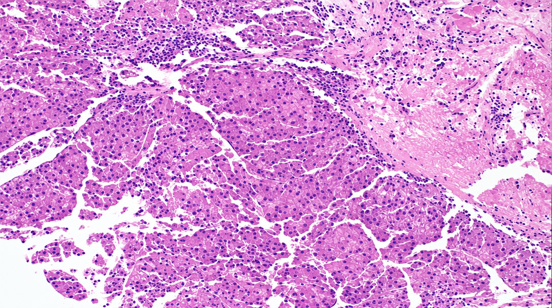
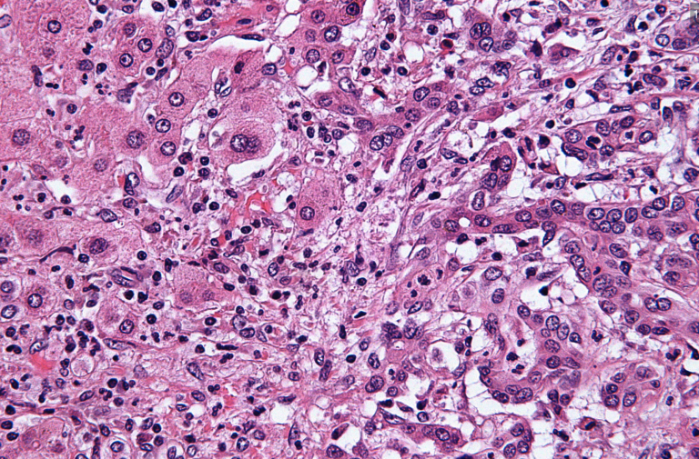
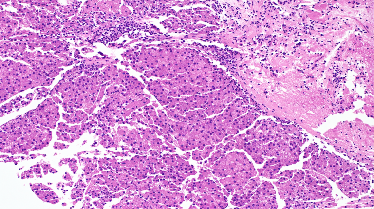
A pathologist orders immunohistochemistry stains when the routine H&E slide leaves an important question open. On a liver biopsy, that question may be whether a mass is hepatocellular carcinoma, cholangiocarcinoma, metastatic cancer from another organ, a benign liver lesion, or unusual reactive tissue. The microscope gives essential clues, but some tumors overlap in appearance. Arginase-1 HepPar-1 Glypican-3 helps narrow the possibilities by testing whether the tissue shows hepatocellular differentiation.
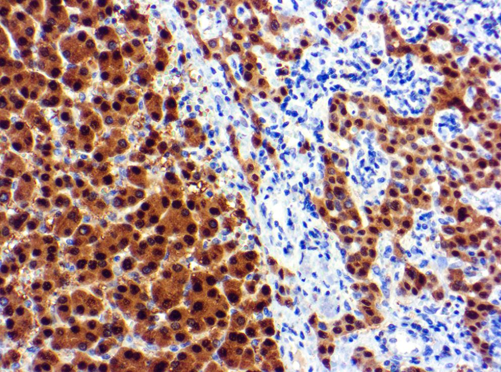
Arginase-1 is often considered a sensitive and specific marker for hepatocellular differentiation, although no marker is perfect. HepPar-1 also supports liver-cell origin but may stain some non-liver tumors in certain settings. Glypican-3 can support hepatocellular carcinoma when the microscopic pattern fits, but it must be interpreted carefully because occasional other tumors can show expression. In other words, immunohistochemistry stains are evidence, not an automatic verdict.
The reason these liver biopsy markers appear together is that pathologists generally prefer a pattern-based approach. A malignant liver tumor may need a broad panel that includes markers for liver origin, bile duct origin, gastrointestinal origin, lung origin, breast origin, melanoma, or other possibilities, depending on the clinical situation. The pathology report may also mention background cirrhosis, inflammation, fatty change, necrosis, vascular invasion, or tumor grade. Those details can change the meaning of a liver tumor diagnosis as much as the stains themselves.
How Serious Are Arginase-1 HepPar-1 Glypican-3 Results?
Arginase-1 HepPar-1 Glypican-3 results can be serious when they support hepatocellular carcinoma or another malignant liver tumor, but the seriousness depends on the entire report. A positive marker panel is not the same as a complete cancer stage. Staging usually requires imaging, tumor size, number of lesions, blood vessel involvement, liver function, and sometimes additional laboratory results such as alpha-fetoprotein. The biopsy answers the tissue-identity question; the clinical team answers the whole-patient question.
In many liver biopsies, Arginase-1 HepPar-1 Glypican-3 is used because the pathologist already sees a concerning growth pattern. Strong Arginase-1 with supportive HepPar-1 and Glypican-3 may strengthen the diagnosis of hepatocellular carcinoma, especially when the tumor architecture and clinical findings match. However, a marker that is negative, weak, or only focal does not always rule out cancer. Small biopsies can sample only part of a tumor, and some cancers have unusual staining patterns.
The emotional weight of this result is real. Words such as positive and carcinoma can make the report feel final before a patient has had time to breathe. A careful pathology review can separate what is confirmed from what is still being evaluated. When the diagnosis is unexpected, when the biopsy is limited, or when treatment will be major, a second review of the slides may be reasonable; the article on when to get a second pathology opinion explains situations where review is especially helpful.
What Happens Next: Treatment And Monitoring
After these liver biopsy markers are reported, the next step is usually clinicopathologic correlation. That phrase means the treating team compares the pathology report with imaging, blood work, liver disease history, viral hepatitis status, alcohol-related liver injury, metabolic liver disease, and overall health. If the diagnosis is hepatocellular carcinoma, the case may be discussed by a multidisciplinary tumor board. Surgeons, hepatologists, oncologists, radiologists, and pathologists may all contribute to the liver tumor diagnosis.
Treatment is not chosen from Arginase-1 HepPar-1 Glypican-3 alone. Options may include surgery, ablation, embolization, radiation-based liver-directed therapy, transplant evaluation, systemic therapy, or close monitoring in selected situations. Liver function can be as important as tumor size because the liver must be strong enough to tolerate certain treatments. This is one reason the background liver in the biopsy can matter, not only the tumor.
Monitoring may involve repeat imaging, blood tests, liver function tests, and sometimes additional biopsy or molecular testing. Immunohistochemistry stains may also be repeated or expanded if the first panel is incomplete or if a second institution reviews the case. Patients who want to understand the structure of the diagnosis, comment, and stain sections may find guidance on reading a pathology report useful before the oncology or hepatology visit. A calm review often makes the next appointment more productive.
Questions To Ask Your Doctor Or Pathologist
- Does the staining pattern support hepatocellular carcinoma, or is the diagnosis still uncertain?
- Were Arginase-1, HepPar-1, and Glypican-3 all positive, or were some stains weak, focal, or negative?
- Does the biopsy show hepatocellular differentiation, bile duct differentiation, or features of a metastatic tumor?
- Was the biopsy sample large enough to make a confident liver tumor diagnosis?
- Does the background liver show cirrhosis, hepatitis, fatty liver disease, or other injury?
- Were additional immunohistochemistry stains performed to rule out cancer from another organ?
- Would a second pathology opinion change treatment planning or confirm eligibility for a specific therapy?
These questions are not meant to challenge the care team. They help translate technical findings into decisions that affect treatment, staging, and follow-up. A patient can ask whether the final diagnosis is definite, favored, suspicious, or pending additional studies, because those words carry different levels of certainty.
If a major treatment decision is being considered, expert slide review can be valuable. Instructions for obtaining a review are outlined in how to get a second opinion on a pathology diagnosis. A second opinion typically reviews the original slides, blocks if available, stains, report wording, and clinical context.
Frequently Asked Questions
Does Arginase-1 HepPar-1 Glypican-3 mean liver cancer?
Arginase-1 HepPar-1 Glypican-3 can support a diagnosis of hepatocellular carcinoma, but the stains alone do not equal the entire diagnosis. The pathologist also evaluates the cell shape, tissue architecture, growth pattern, and background liver. In some reports, the markers provide strong confirmation; in others, they are one part of a broader workup. The final diagnosis line is more important than any single stain name.
What does Glypican-3 positive mean on a liver biopsy?
Glypican-3 positive means the tested cells show staining for a protein that is often seen in hepatocellular carcinoma. It can be helpful when the pathologist is separating cancer from benign liver nodules or from some other tumors. However, Glypican-3 is not interpreted by itself. The result has the most meaning when compared with Arginase-1, HepPar-1, and the microscopic appearance.
Is Arginase-1 more accurate than HepPar-1?
Arginase-1 is often considered very useful for showing hepatocellular differentiation and may be more specific than HepPar-1 in some diagnostic settings. HepPar-1 remains helpful, especially when used as part of a panel. Neither stain is perfect in every case. Pathologists often rely on the combined pattern of liver biopsy markers rather than choosing one winner.
Can a second opinion change a liver biopsy diagnosis?
A second opinion can change, refine, or confirm a liver biopsy diagnosis, especially when the sample is small, the tumor is unusual, or treatment decisions are high stakes. Arginase-1 HepPar-1 Glypican-3 may be reviewed along with the original slides and any additional immunohistochemistry stains. Sometimes the diagnosis stays the same, but the level of confidence becomes clearer. That clarity can still be very valuable for treatment planning.
Do these markers show the stage of liver cancer?
No. These markers help identify the type and origin of the tumor, but they do not provide the cancer stage by themselves. Staging usually depends on imaging, tumor size, number of tumors, vascular invasion, spread outside the liver, and liver function. The pathology report may provide important pieces of the staging puzzle, but the full stage comes from combining several sources of information.
Arginase-1 HepPar-1 Glypican-3 results are technical, but they are not meant to leave patients in the dark. They are tools pathologists use to identify what a liver biopsy is showing and how confident the diagnosis can be. Honest Pathology consultations can help patients and families understand whether the wording of a report is definite, uncertain, or worth further expert review.
References:
National Cancer Institute — Pathology Reports Fact Sheet
National Cancer Institute — Definition of Hepatocellular Carcinoma
NCBI Bookshelf — Biomedical Literature And Clinical References