Scared by a testicular cancer report? seminoma vs nonseminoma affects treatment choices. Learn what the terms mean and what to ask next.
Seminoma vs nonseminoma is one of the most important distinctions in a testicular cancer pathology report because it helps guide staging, treatment, and follow-up. Both are types of germ cell tumor, but they can behave differently and respond to treatment in different ways. The wording may look technical, especially when the report includes terms such as embryonal carcinoma, yolk sac tumor, teratoma, choriocarcinoma, lymphovascular invasion, or rete testis involvement.
Seeing a cancer diagnosis in writing can make time feel as if it has stopped. Many patients and families read the same paragraph repeatedly, searching for the one sentence that explains what will happen next. A pathology report is not meant to be frightening, but it is written for doctors, which can make it feel cold and confusing. Clear interpretation can turn that report from a source of panic into a practical map for the next conversation.
Seminoma Vs Nonseminoma — What It Actually Means
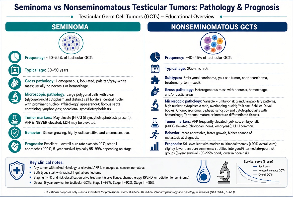
Seminoma vs nonseminoma describes two broad categories of testicular cancer that arise from germ cells, the cells involved in sperm production. Seminoma is usually made of one tumor type with a fairly uniform microscopic appearance. Nonseminoma is a broader group that may include embryonal carcinoma, yolk sac tumor, choriocarcinoma, teratoma, or a mixture of several components. A mixed germ cell tumor is usually treated as nonseminoma if any nonseminomatous component is present.
A helpful analogy is to think of testicular cancer as a family name and seminoma or nonseminoma as different branches of that family. The shared family origin matters, but the branch tells the oncology team how the tumor may behave. Seminoma often grows and spreads in a more predictable pattern and is highly sensitive to radiation and chemotherapy. Nonseminoma can sometimes grow more quickly, spread earlier, or produce specific tumor markers that help with monitoring.
Pathologists make this distinction by examining tissue from an orchiectomy or biopsy under the microscope, then matching the appearance with special tests when needed. A pathology report may describe the percentage of each component, especially when the tumor is mixed. For patients trying to understand the basic structure of a report, What Is a Pathology Report? explains how these documents are organized. In this setting, testicular cancer, germ cell tumor, pathology report, and tumor markers are all linked pieces of the same diagnostic puzzle.
Why Your Report Shows This Finding
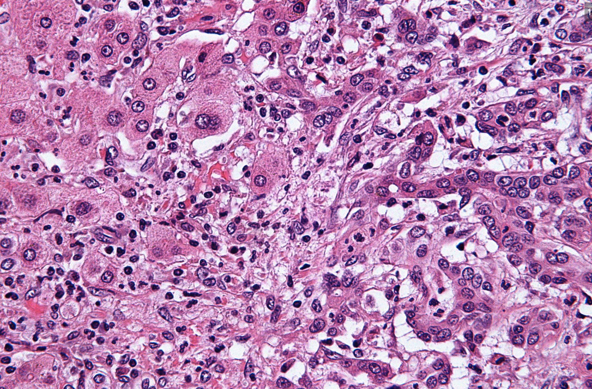
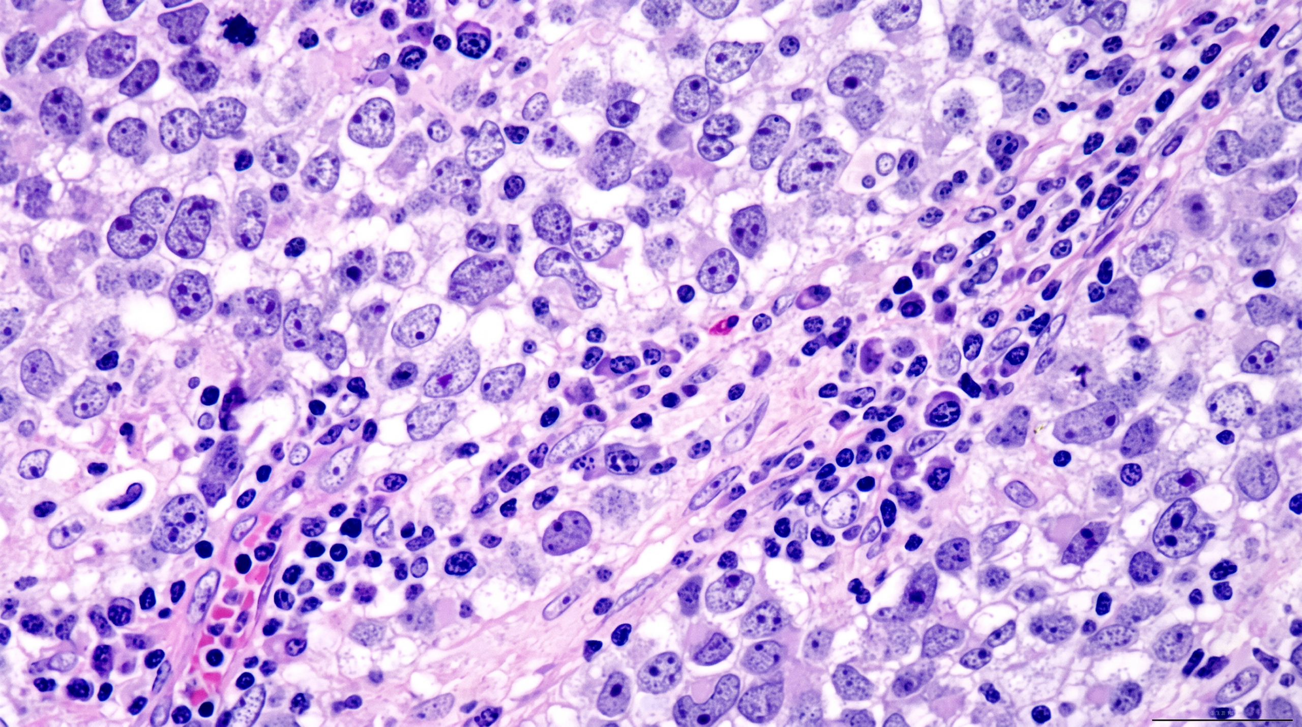
A report shows seminoma vs nonseminoma because the pathologist sees features that place the tumor into one or both categories. Seminoma cells often look relatively uniform, with clear cytoplasm and a background of lymphocytes. Nonseminomatous areas may look more varied, with gland-like patterns, primitive solid growth, cystic spaces, hemorrhage, necrosis, or tissue resembling teratoma. Embryonal carcinoma is especially important because even a small amount can influence treatment planning.
The pathology report may also mention lymphovascular invasion, which means cancer cells are seen inside small blood vessels or lymphatic channels. This does not automatically mean distant spread has occurred, but it can raise the risk that cancer cells have moved beyond the testicle. Rete testis involvement may also be described, especially in seminoma, because it can contribute to risk assessment in some clinical settings. These findings are not moral judgments or signs that anything was missed; they are microscopic observations that help estimate risk.
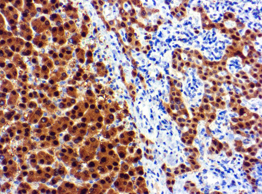
Tumor markers are another reason the distinction matters. Blood tests such as AFP, beta-hCG, and LDH are often checked before and after surgery. AFP is generally not produced by pure seminoma, so an elevated AFP level usually suggests a nonseminomatous component, even if the sampled tissue looks like seminoma. The oncology team combines tumor markers, imaging, physical examination, and the pathology report to determine stage and treatment options.
How Serious Is Seminoma Vs Nonseminoma?
Seminoma vs nonseminoma can sound like a verdict, but it is better understood as a treatment guide. Testicular cancer is often highly treatable, even when it has spread, and cure rates are very high compared with many other cancers. The seriousness depends on stage, tumor markers, imaging findings, lymphovascular invasion, and the exact mixture of tumor types. A small, localized seminoma without high-risk features is a very different situation from a nonseminomatous germ cell tumor with high markers and enlarged lymph nodes.
For seminoma, many patients with disease limited to the testicle may be managed with surveillance, chemotherapy, or radiation depending on stage and risk factors. Rete testis involvement and tumor size may be discussed, although treatment decisions vary by guideline and clinical context. For nonseminoma, the presence of embryonal carcinoma, lymphovascular invasion, and marker behavior can influence whether surveillance, chemotherapy, or retroperitoneal lymph node dissection is considered. Seminoma vs nonseminoma matters because the same stage number may lead to different recommended paths.
The report should be read as part of the full clinical picture, not as a single frightening phrase. Tumor markers that fall appropriately after surgery can be reassuring, while persistently elevated tumor markers may prompt more evaluation. Imaging studies look for lymph node or organ involvement, but small microscopic spread cannot always be seen on a scan. A patient-friendly overview such as Understanding Your Pathology Report: How to Read It with Confidence can help families connect the diagnosis line, stage-related findings, and next-step questions.
What Happens Next: Treatment And Monitoring
After a diagnosis of seminoma vs nonseminoma, the next step is usually a review of the complete pathology report, tumor markers, and imaging. The oncology or urology team typically confirms the stage, checks whether markers normalized after orchiectomy, and explains treatment choices. Some patients need active surveillance with scheduled blood tests and scans. Others may need chemotherapy, radiation therapy, surgery to remove lymph nodes, or a combination of approaches.
Monitoring is not passive; it is an organized plan designed to catch recurrence early if it occurs. Tumor markers may be checked repeatedly because changes can appear before symptoms develop. Imaging schedules depend on the tumor type, stage, prior treatment, and current guidelines. For testicular cancer, follow-up is especially important during the first few years, when the risk of recurrence is highest.
A second pathology review may be useful when the diagnosis is mixed, the percentages are unclear, the report conflicts with tumor markers, or treatment options would change based on the exact classification. This is particularly relevant for a germ cell tumor with unusual features or a small nonseminomatous component. When Should You Get a Second Pathology Opinion? explains situations where review can add clarity. A second review does not mean the first pathologist made an error; it means the tissue is important enough to deserve the highest level of certainty.
Questions To Ask Your Doctor Or Pathologist
- Does the pathology report diagnose pure seminoma, pure nonseminoma, or a mixed germ cell tumor?
- What percentage of each tumor component is present, including embryonal carcinoma, yolk sac tumor, teratoma, or choriocarcinoma?
- Is lymphovascular invasion present, absent, or indeterminate?
- Does the report mention rete testis involvement, tunica invasion, spermatic cord involvement, or margin status?
- Do the tumor markers match the diagnosis, especially AFP, beta-hCG, and LDH?
- What stage is the testicular cancer based on the pathology report, imaging, and blood tests?
- Would a second pathology opinion change surveillance, chemotherapy, radiation, or surgery recommendations?
These questions help turn a difficult report into a focused discussion. The goal is not to memorize pathology language, but to understand which findings change care. If the diagnosis includes seminoma vs nonseminoma, the exact wording can matter because treatment pathways are not always identical.
Patients and families can also ask whether the slides have been reviewed by a pathologist with experience in genitourinary tumors. For complex or mixed cases, How to Get a Second Opinion on Your Pathology Diagnosis can help explain the practical steps. A careful review can confirm the diagnosis, clarify tumor percentages, and support confident treatment decisions.
Frequently Asked Questions
Is seminoma better than nonseminoma?
Seminoma is often slower growing and very sensitive to treatment, but “better” is not the right way to frame the diagnosis. Many nonseminomatous tumors are also highly curable, especially when found early and treated appropriately. Seminoma vs nonseminoma helps doctors choose the safest and most effective treatment plan. Stage, tumor markers, and lymphovascular invasion often matter as much as the name itself.
Can a testicular tumor be both seminoma and nonseminoma?
Yes. A mixed germ cell tumor can contain seminoma plus one or more nonseminomatous components. When any nonseminomatous component is present, treatment is often planned as nonseminoma. The pathology report should list the components and, when possible, the approximate percentage of each.
Does AFP mean nonseminoma?
AFP is a tumor marker that is not expected to be elevated in pure seminoma. If AFP is elevated, doctors usually look carefully for a nonseminomatous component, even if the tissue sample looks mostly like seminoma. This is one reason tumor markers and the pathology report must be interpreted together. Seminoma vs nonseminoma may be reconsidered if the blood tests and microscopic findings do not fit.
What does lymphovascular invasion mean in testicular cancer?
Lymphovascular invasion means cancer cells are seen inside small vessels or lymphatic channels under the microscope. This finding can increase the risk that cancer cells have traveled beyond the testicle. It does not prove that cancer has spread to distant organs. It is one factor used with stage, imaging, and tumor markers to decide follow-up or treatment.
Should I get a second opinion on a germ cell tumor pathology report?
A second opinion can be helpful when the diagnosis is mixed, rare, unexpected, or likely to affect treatment choices. Germ cell tumor classification can be straightforward in many cases, but small components such as embryonal carcinoma may change management. A review may also help when tumor markers do not match the diagnosis. For seminoma vs nonseminoma, confirmation can provide practical reassurance before major treatment decisions.
A testicular cancer pathology report can feel overwhelming, but each term has a purpose. The distinction between seminoma vs nonseminoma is meant to guide care, not define the whole future. Honest Pathology consultations can help clarify difficult wording, explain why specific findings matter, and support more confident conversations with the treating team.
References:
National Cancer Institute — Testicular Cancer
National Cancer Institute — Pathology Reports Fact Sheet
National Cancer Institute — Germ Cell Tumor Definition