Worried by rete testis invasion on a report? Learn what it means, why it is documented, and what to ask the cancer team next.
Rete testis invasion means a testicular tumor has grown into a small drainage-network area inside the testis, and pathologists document it because it can help complete the overall risk picture. This phrase is most often seen on an orchiectomy pathology report after removal of a testicle for suspected or confirmed testicular cancer. It does not automatically mean that cancer has spread throughout the body, and it is only one part of testicular cancer staging and follow-up planning.
Seeing unfamiliar anatomy in a cancer report can feel frightening, especially when the wording sounds technical and final. Many patients read the report before a full explanation from the treating clinician, which can make a single line feel larger than it is. Clear context matters because pathology reports are built like evidence files: each finding is recorded so the care team can make the safest plan.
A helpful starting point is that rete testis invasion is a local microscopic finding, not a diagnosis by itself. It has to be interpreted with the tumor type, tumor size, lymphovascular invasion, imaging results, blood tumor markers, and the clinical situation.
Rete Testis Invasion — What It Actually Means
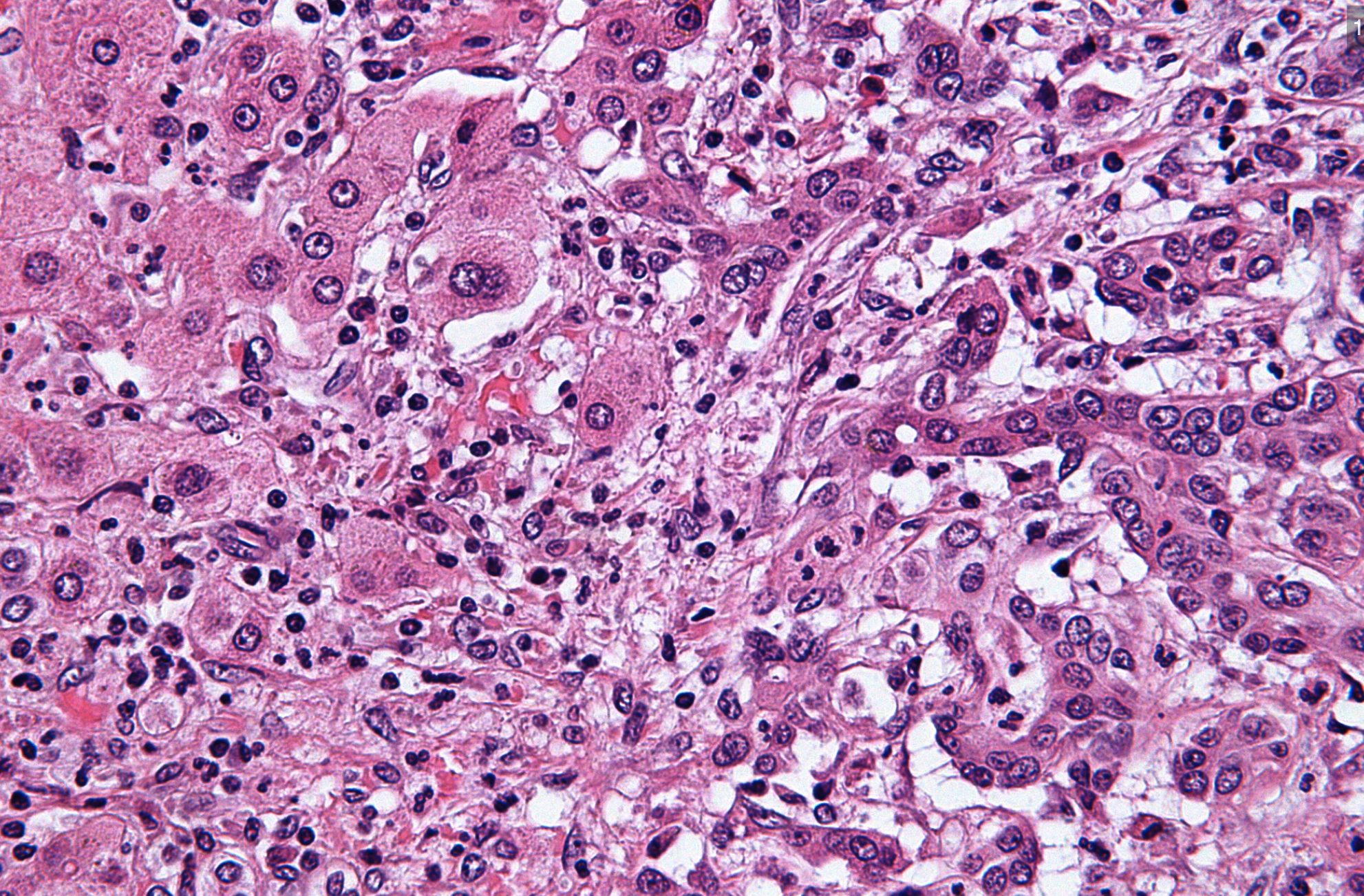
The rete testis is a small network of channels located near the back of the testis, close to the area where sperm normally drains toward the epididymis. In simple terms, it works like a small collection hub inside the testicle. Rete testis invasion is documented when tumor cells are seen growing into that area under the microscope. Pathologists include it because the location of tumor growth can add useful information to an orchiectomy pathology report.
A real-world analogy is a water system in a building. If a problem begins in one room, the report may document whether it has reached a hallway, a pipe, or a connecting space. That does not necessarily mean the problem has reached the whole building, but it describes the path and extent of local involvement. In the same way, rete testis invasion describes local tumor extension within the testis and nearby internal structures.
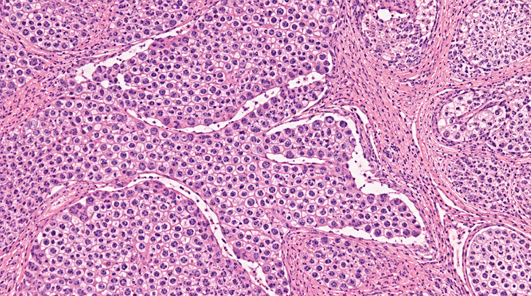
This finding is especially discussed in seminoma, one major type of germ cell tumor. In some clinical guidelines and studies, rete testis invasion has been evaluated as one of several seminoma risk factors for relapse after orchiectomy, particularly in stage I disease. However, the significance of this feature is not the same for every patient or every tumor type. The final meaning depends on the entire pathology report, not on this line alone. For patients learning the structure of a report, What Is a Pathology Report? can help explain why pathologists list detailed anatomic findings.
Why Your Report Shows This Finding
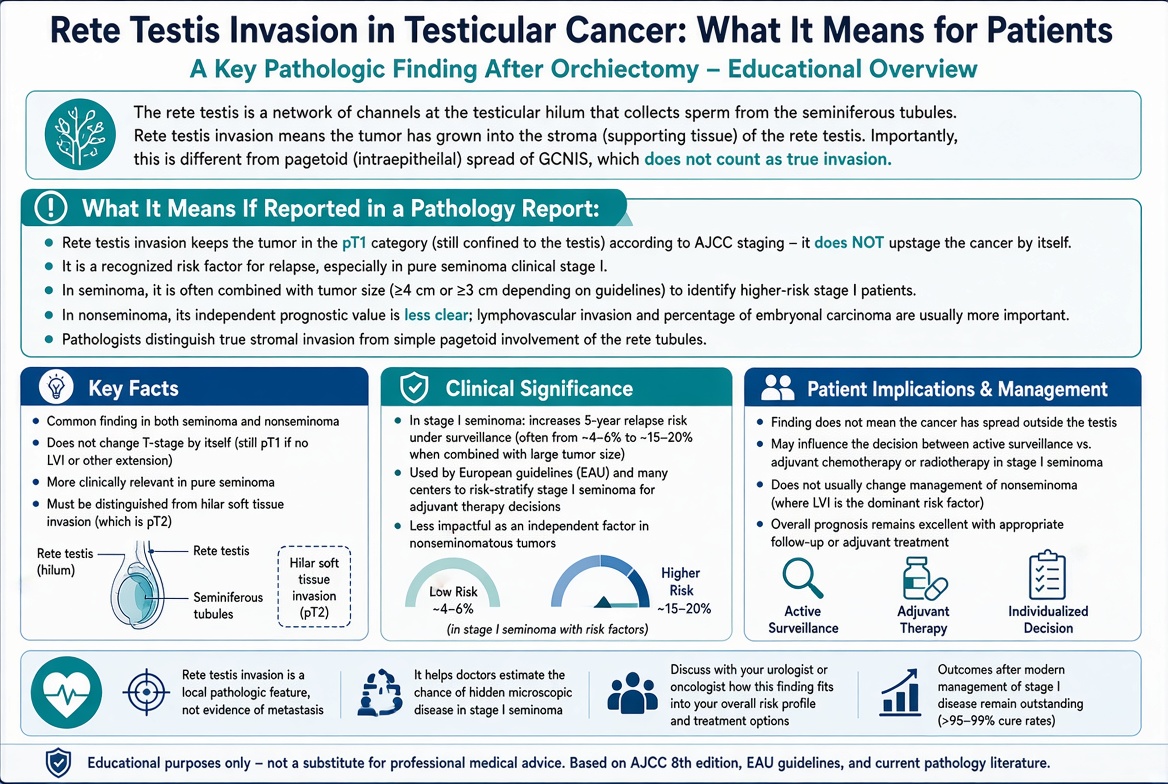
An orchiectomy pathology report usually documents tumor type, tumor size, margins, lymphovascular invasion, spermatic cord involvement, rete testis involvement, and whether tumor extends beyond the testis. These elements help clinicians understand the behavior of a germ cell tumor and compare the case with established staging systems. The pathologist examines representative tissue sections under the microscope and records where tumor cells are present. Rete testis invasion appears in the report because it is one of the anatomic sites that may be involved.
The finding can be described in slightly different ways. Some reports may say “invades rete testis,” “involves rete testis,” or “rete testis invasion present.” Other reports may distinguish between pagetoid spread along rete testis lining cells and true stromal invasion, depending on the reporting system and institutional practice. That distinction can matter because not every pattern of microscopic involvement carries the same implication. If wording is unclear, the treating doctor may ask the pathologist to clarify the exact pattern of tumor extension.
Several other findings are usually reviewed at the same time. Lymphovascular invasion means tumor cells are seen in small blood vessels or lymphatic spaces, and it can be an important risk feature in some germ cell tumor types. Spermatic cord involvement is a separate finding and can affect pathologic stage when tumor extends into that structure. Testicular cancer staging also considers whether cancer is limited to the testis, involves adjacent structures, or has evidence of spread on imaging or tumor marker testing. A patient trying to understand multiple lines in a report may find Understanding Your Pathology Report: How to Read It with Confidence useful before the oncology visit.
How Serious Is Rete Testis Invasion?
Rete testis invasion can be clinically relevant, but it is not automatically a sign of advanced cancer. In many patients, the tumor is still considered localized after orchiectomy, especially when imaging is negative and blood tumor markers are appropriate. The seriousness depends on the tumor type, stage, tumor size, lymphovascular invasion, and whether any disease is seen outside the testis. Rete testis invasion is one piece of a larger puzzle.
For seminoma, rete testis invasion has historically been studied as one of the seminoma risk factors associated with recurrence risk in stage I disease. Some studies found that larger tumor size and rete testis invasion may be associated with higher relapse risk, while other analyses have questioned how strongly these features should guide treatment by themselves. Because of that, many clinicians use this finding as part of a discussion rather than as a single deciding factor. Testicular cancer staging and surveillance planning usually rely on a combination of pathology, imaging, blood tests, and patient preferences.
For nonseminomatous germ cell tumor, other features, especially lymphovascular invasion and the percentage or presence of certain tumor components, may carry more weight in risk assessment. Rete testis invasion may still be documented because it describes tumor extension and supports accurate reporting. A report showing rete testis invasion but no spermatic cord involvement, negative margins, and no radiologic spread may lead to a very different plan than a report with multiple higher-risk features. The most useful question is not simply whether the phrase is present, but how it changes the patient’s actual recurrence risk and follow-up options.
What Happens Next: Treatment And Monitoring
After an orchiectomy pathology report is finalized, the care team usually combines the microscopic findings with blood tumor markers such as AFP, beta-hCG, and LDH, as well as CT imaging when appropriate. This combined review helps determine testicular cancer staging and whether the next step is surveillance, chemotherapy, radiation therapy, surgery, or another approach. In stage I seminoma, management may include surveillance, adjuvant therapy, or individualized discussion based on risk features and patient priorities. Rete testis invasion may be part of that conversation, but it does not replace the rest of the staging workup.
Monitoring often includes scheduled physical exams, blood tests, and imaging at defined intervals. The exact schedule varies by tumor type, stage, treatment choice, and national or institutional guideline. Surveillance can feel emotionally difficult because it asks patients to live with uncertainty, but it is a deliberate medical strategy in selected cases. The purpose is to detect recurrence early, when treatment is usually highly effective.
A second pathology review may be helpful when the tumor type is mixed, the report wording is unclear, or treatment decisions depend heavily on subtle microscopic features. This is especially true for a germ cell tumor with several components or borderline patterns of tumor extension. A second opinion does not mean the first pathologist was wrong; it is a safety check before major decisions. Patients considering review can read When Should You Get a Second Pathology Opinion? for practical context.
Questions To Ask Your Doctor Or Pathologist
- Does rete testis invasion change the pathologic stage in this specific case?
- Is the tumor seminoma, nonseminomatous germ cell tumor, or a mixed germ cell tumor?
- Were lymphovascular invasion, spermatic cord involvement, or positive margins present?
- What was the tumor size, and does it affect recurrence-risk estimates?
- Do the blood tumor markers match the pathology findings?
- What surveillance or treatment options are being considered, and why?
- Would a second pathology review change any treatment recommendation?
These questions can help turn an intimidating orchiectomy pathology report into a more organized discussion. A report is not meant to be read as isolated phrases; it is meant to be interpreted as a connected medical document. Rete testis invasion should be discussed in relation to the diagnosis, stage, imaging, and tumor markers.
When the report contains complex wording, a pathology consultation can clarify what was actually seen under the microscope. If slides are reviewed elsewhere, the reviewing pathologist may confirm the diagnosis, refine the subtype, or clarify features such as tumor extension. Patients who need practical steps can use How to Get a Second Opinion on Your Pathology Diagnosis as a starting framework.
Frequently Asked Questions
Does rete testis invasion mean cancer has spread?
Rete testis invasion does not automatically mean that cancer has spread to lymph nodes or distant organs. It means tumor cells involve a specific local structure within the testis. Spread outside the testis is assessed with staging information, imaging, blood tumor markers, and other pathology findings. The cancer team can explain whether the overall case is localized or more advanced.
Is rete testis invasion common in seminoma?
Rete testis invasion can be seen in seminoma and is often specifically documented because it has been studied as one of the seminoma risk factors. Its meaning varies depending on tumor size, imaging, tumor markers, and the rest of the report. Some patients with this finding still have stage I disease after orchiectomy. The finding should be interpreted as part of the full clinical picture.
Can rete testis invasion change treatment?
Rete testis invasion may influence treatment discussions in some cases, particularly stage I seminoma, but it rarely acts alone. Treatment decisions usually include testicular cancer staging, tumor size, blood marker results, imaging findings, and patient preferences. Some patients may still be offered surveillance. Others may discuss adjuvant treatment if the full risk profile supports that option.
What is the difference between rete testis invasion and lymphovascular invasion?
Rete testis invasion describes tumor involvement of the rete testis, a small anatomic drainage area inside the testis. Lymphovascular invasion means tumor cells are seen inside small vessels or lymphatic spaces. These are different findings, and they may carry different implications depending on tumor type. Both may appear on an orchiectomy pathology report because both help describe how the tumor is behaving locally.
Should I get a second opinion for rete testis invasion?
A second opinion can be reasonable when the diagnosis is mixed, the wording is unclear, or treatment choices depend on subtle pathology features. Rete testis invasion is usually confirmable on slide review if the relevant tissue sections are available. A second review may also clarify tumor type, margins, lymphovascular invasion, and tumor extension. This can provide reassurance before choosing surveillance or additional treatment.
Rete testis invasion can look alarming on paper, but it is best understood as one documented feature in a larger evidence-based assessment. The most reassuring next step is a clear explanation of how this finding fits with the tumor type, stage, markers, and imaging. Honest Pathology consultations can help patients and families translate complex report language into focused questions for the treating team.
References:
National Cancer Institute — Pathology Reports Fact Sheet
National Cancer Institute — Testicular Cancer
National Cancer Institute Dictionary of Cancer Terms — Germ Cell Tumor