A scary flow cytometry result can raise lymphoma fears. monoclonal B-cell population has several meanings; learn what to ask next.
monoclonal B-cell population means a group of B lymphocytes looks genetically related on testing, but it does not automatically mean lymphoma. This finding is most often reported after flow cytometry, a laboratory test that studies markers on the surface of blood, bone marrow, lymph node, or body fluid cells. The meaning depends on the sample type, the number of abnormal cells, the patient’s age, symptoms, physical exam, blood counts, imaging, and sometimes a lymph node biopsy.
Seeing the word “monoclonal” can feel frightening because it sounds final. In pathology, however, monoclonal describes a pattern, not a complete diagnosis by itself. A monoclonal B-cell population is like seeing a group of people wearing the same uniform in a crowd: The finding deserves attention, but the setting determines whether it represents a small harmless group, an early condition, or a lymphoma that needs treatment. Clear questions and careful correlation often turn panic into a practical plan.
Monoclonal B-Cell Population — What It Actually Means
A monoclonal B-cell population is identified when B cells share the same immunologic signature, most often the same light chain pattern and a similar set of surface markers. Normal B cells are diverse, like many different keys on a key ring. A B-cell clone is more uniform, like many copies of the same key. Flow cytometry can detect that uniformity with high sensitivity, even when the abnormal cells are too few to see clearly under a microscope.
The word “B-cell” refers to one type of white blood cell involved in immune defense and antibody production. In routine practice, pathologists compare the abnormal cells with normal B cells and with known patterns seen in blood and lymph node diseases. Light chain restriction, meaning a strong predominance of either kappa or lambda light chain, is one major clue that a B-cell clone is present. The finding is then interpreted alongside the blood smear, tissue architecture, and clinical information.
A monoclonal B-cell population may be found in several settings. It can appear in chronic lymphocytic leukemia, some low-grade lymphomas, mantle cell lymphoma, marginal zone lymphoma, follicular lymphoma, or other B-cell lymphoproliferative disorders. It can also be very small and found incidentally, especially in blood samples from older adults. For patients trying to understand the larger report, a step-by-step resource such as Understanding Your Pathology Report can help explain how individual findings fit together.
Why The Report Shows This Finding
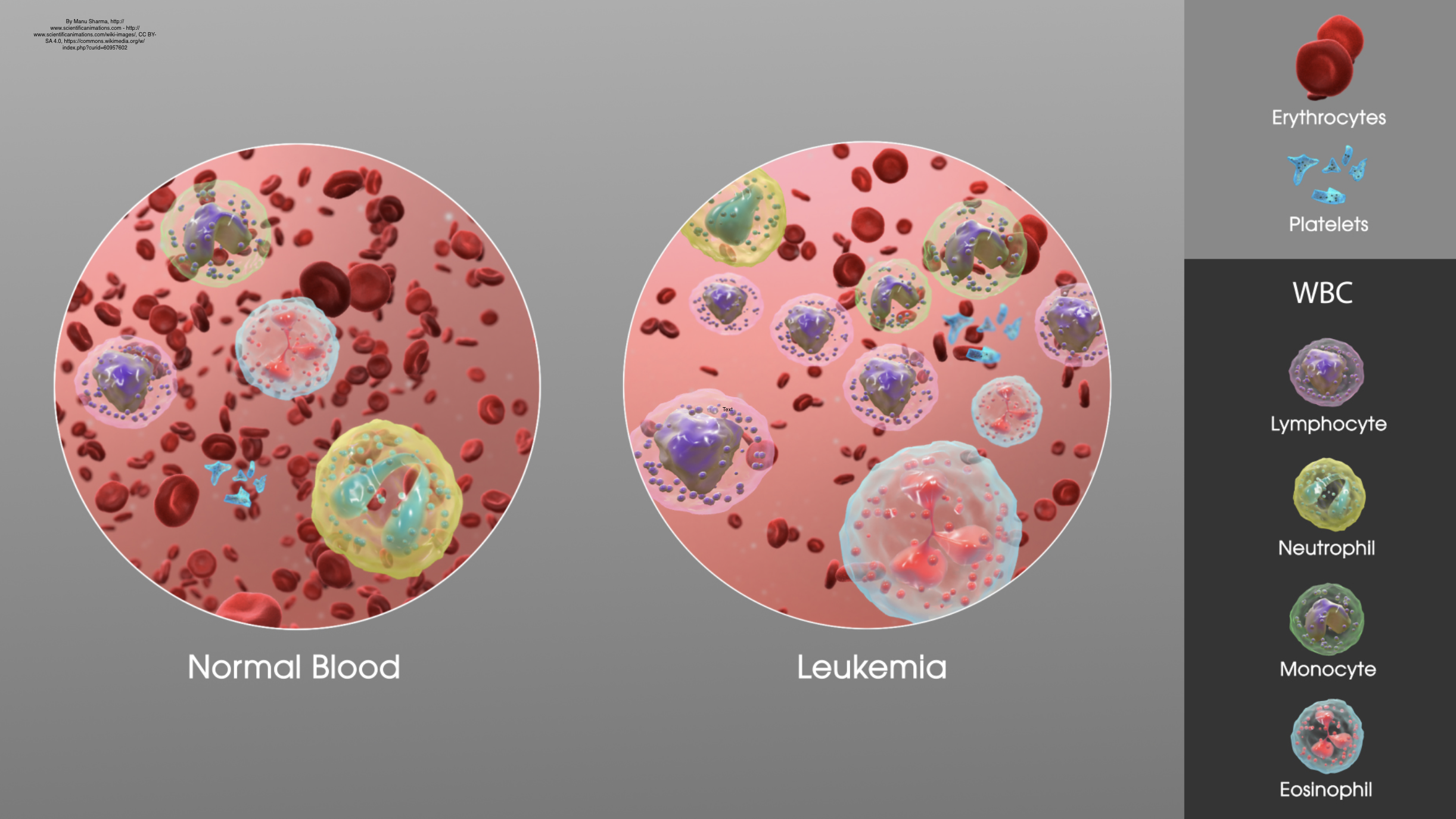
Reports usually show this finding because flow cytometry detected a repeated marker pattern among B cells. The laboratory may describe markers such as CD5, CD10, CD19, CD20, CD23, FMC7, kappa, and lambda, depending on the testing panel. These markers help classify the B-cell clone and suggest whether it resembles chronic lymphocytic leukemia, follicular lymphoma, mantle cell lymphoma, or another process. The pattern is helpful, but it is not always enough to make a complete diagnosis.
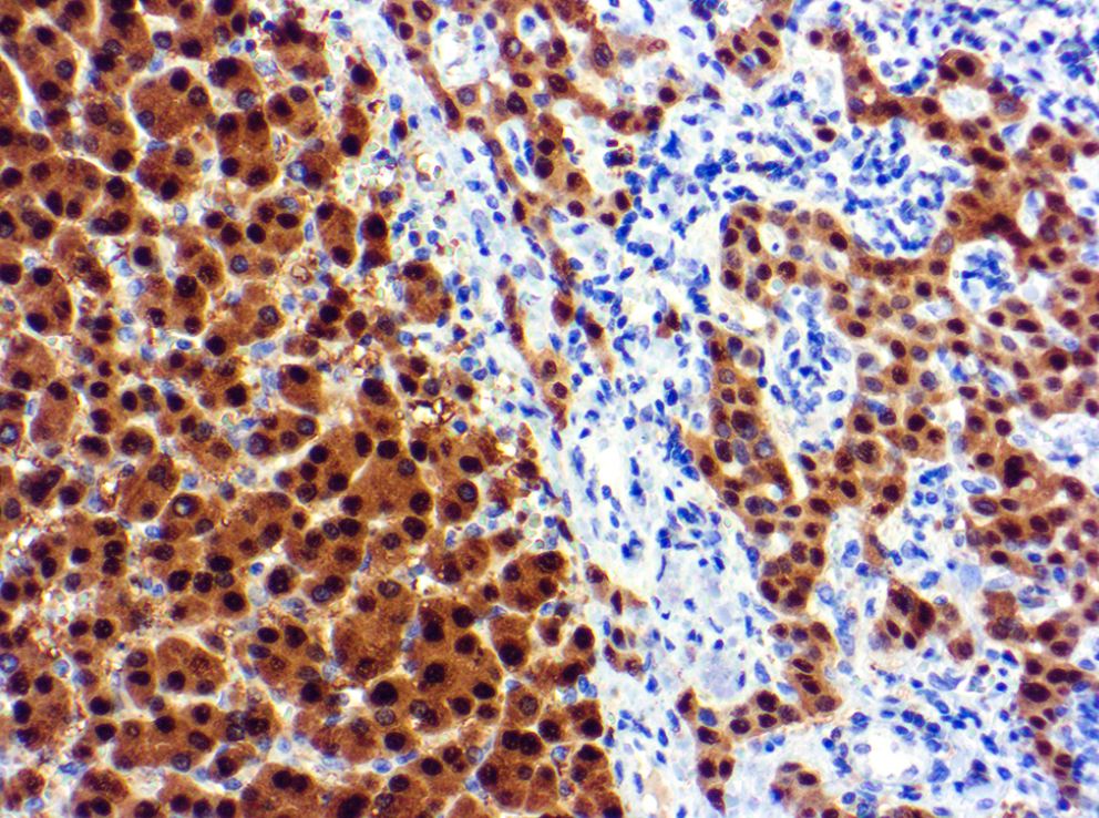
The source of the specimen matters greatly. A small monoclonal B-cell population in peripheral blood can mean something very different from the same finding in an enlarged lymph node biopsy. In blood, the pathologist usually considers the absolute lymphocyte count, the blood smear appearance, and whether the abnormal cells meet criteria for a named disorder. In a lymph node biopsy, tissue structure, cell size, growth pattern, and immunohistochemistry may be needed because architecture can distinguish lymphoma from a tiny background clone.
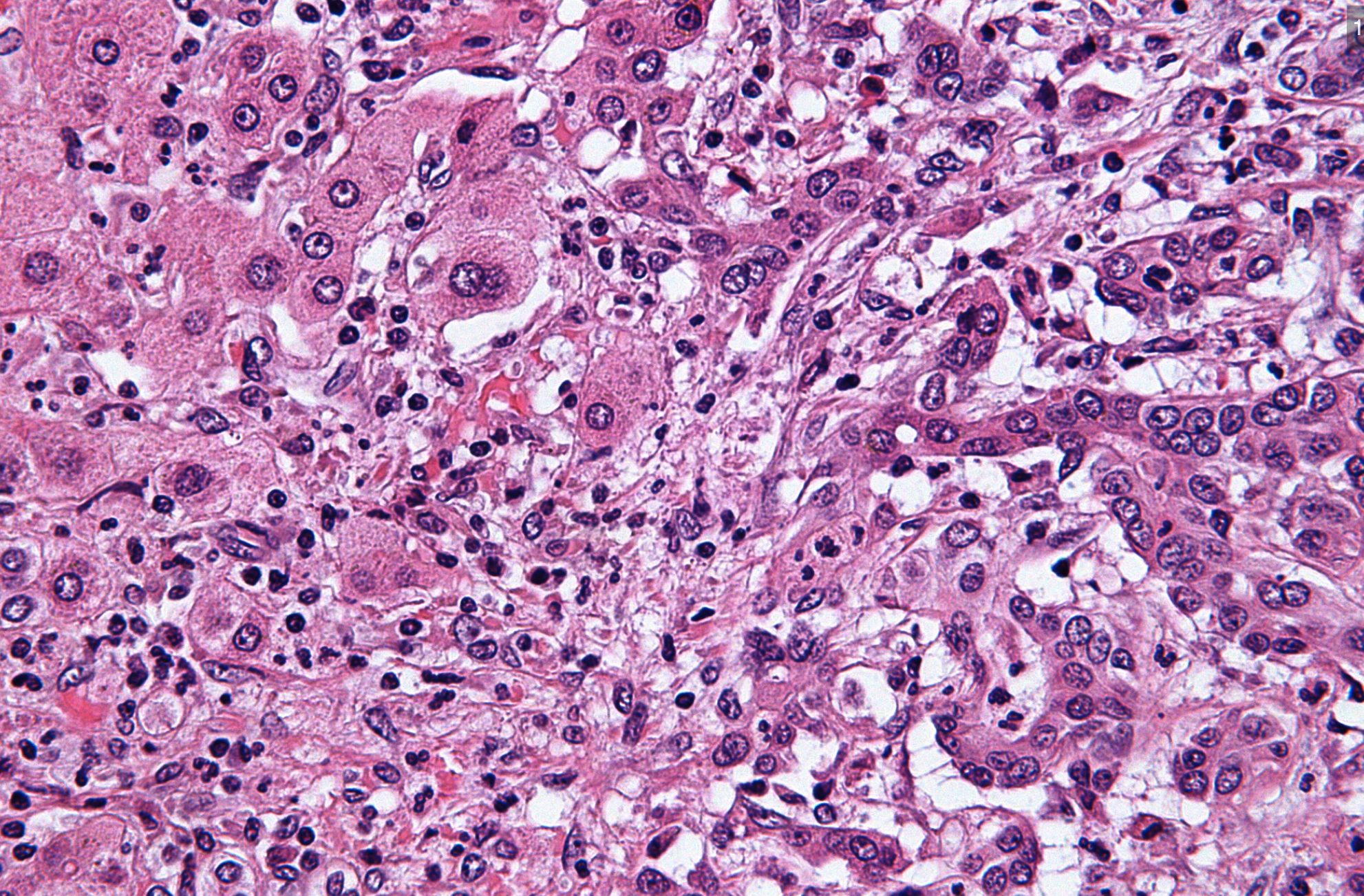
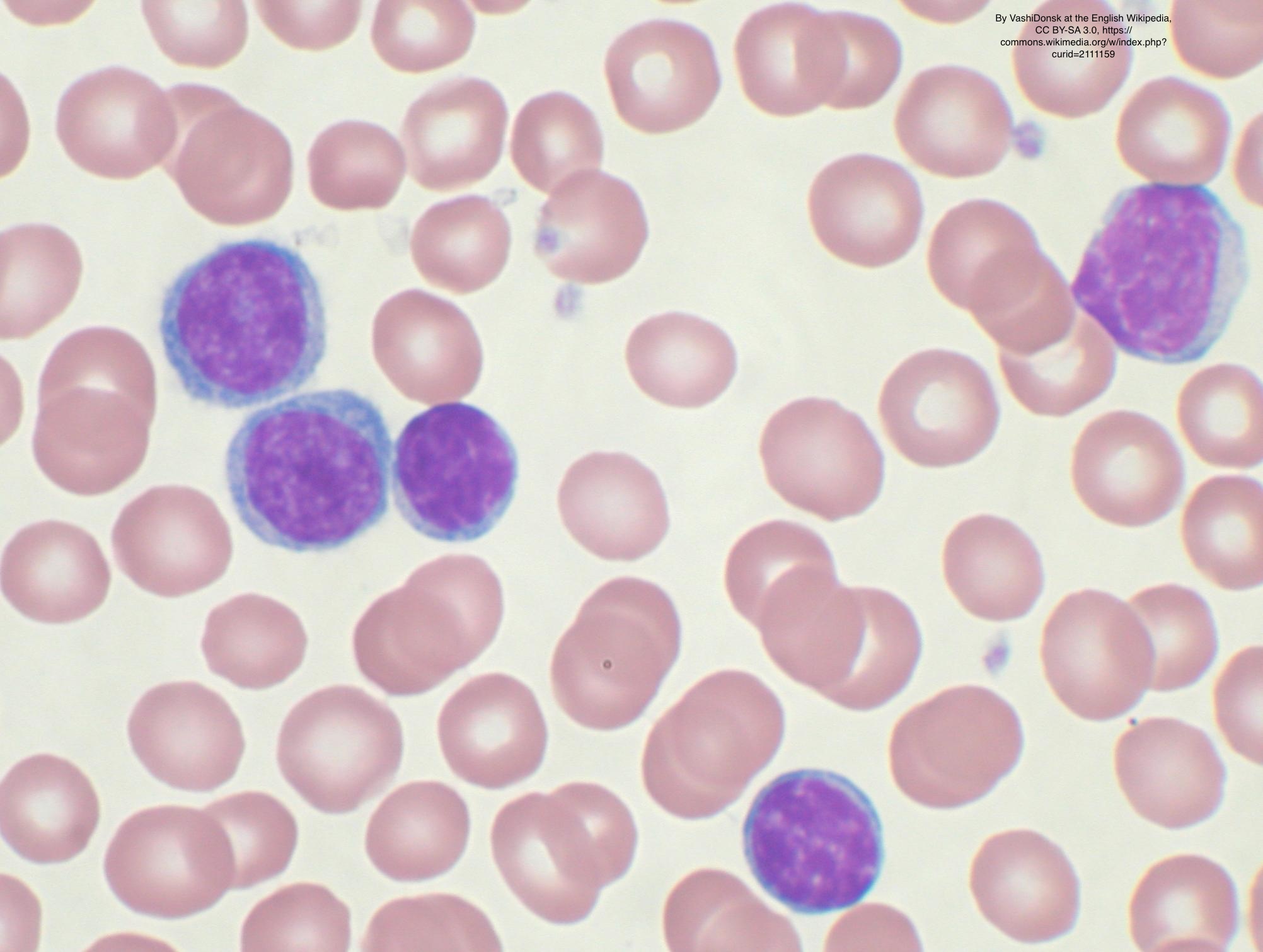
Some monoclonal B-cell findings are incidental. Small B-cell clones may be seen in aging, immune stimulation, autoimmune disease, infections, or after previous treatment for another condition. Light chain restriction supports clonality, but the clinical impact depends on how many cells are involved and where they are found. A blood smear can show whether lymphocytes look mature and small, atypical, large, or numerous, while a lymph node biopsy can show whether normal tissue is replaced by a neoplastic process.
How Serious Is A Monoclonal B-Cell Population?
A monoclonal B-cell population ranges from low-risk and incidental to clinically significant lymphoma, so seriousness cannot be judged from the phrase alone. The most reassuring scenario is a very small B-cell clone in blood, normal blood counts, no enlarged lymph nodes, no enlarged spleen, and no concerning symptoms. In that setting, doctors may use terms such as monoclonal B-cell lymphocytosis if formal criteria are met. Monitoring rather than treatment is common when the clone is small and the patient is well.
A more concerning scenario occurs when the monoclonal B-cell population is large, associated with rising lymphocyte counts, anemia, low platelets, bulky lymph nodes, night sweats, weight loss, fever, or organ involvement. A B-cell clone with certain marker patterns may raise concern for chronic lymphocytic leukemia or mantle cell lymphoma, but classification requires full clinicopathologic correlation. Mantle cell lymphoma, for example, often needs additional testing such as cyclin D1, SOX11, or genetic studies, depending on the specimen. The report may recommend additional studies instead of giving a final label immediately.
The sample type also changes the level of concern. A monoclonal B-cell population in a lymph node biopsy that replaces normal lymph node architecture is more likely to represent lymphoma than a tiny clone in blood found during unrelated testing. A blood smear can support the interpretation by showing whether abnormal lymphocytes are numerous or morphologically suspicious. When the wording feels unclear, a second pathology review can be reasonable, especially before major treatment decisions; patients can learn more at When Should You Get a Second Pathology Opinion?.
What Happens Next: Treatment And Monitoring
The next step is usually not based on flow cytometry alone. Doctors often review the complete blood count, blood smear, physical exam, symptoms, imaging if available, and any tissue biopsy. If the monoclonal B-cell population is small and the patient has no symptoms, repeat blood testing may be recommended after several months. If lymph nodes are enlarged or imaging is abnormal, a lymph node biopsy may be needed to evaluate architecture and confirm the exact diagnosis.
Treatment depends on the final diagnosis, not simply the presence of a B-cell clone. Some patients with chronic lymphocytic leukemia are monitored for years before therapy is needed. Other lymphomas may require treatment sooner, depending on subtype, stage, growth rate, symptoms, and organ involvement. The blood smear and flow cytometry results help guide that pathway, but clinical context remains central.
Additional tests may include immunohistochemistry, FISH, chromosome studies, molecular testing, or Ki-67 proliferation assessment, depending on the suspected disease. Ki-67 is sometimes used in lymphoma workups to estimate how actively cells are dividing; a separate explanation is available at What Does Ki-67 Mean and How High Is Concerning?. If treatment is being considered, confirming the diagnosis before therapy is essential. A carefully reviewed pathology report often prevents both undertreatment and overtreatment.
Questions To Ask The Doctor Or Pathologist
- What percentage and absolute number of cells make up the monoclonal B-cell population?
- Was the B-cell clone found in blood, bone marrow, body fluid, or a lymph node biopsy?
- Does the flow cytometry pattern suggest chronic lymphocytic leukemia, mantle cell lymphoma, follicular lymphoma, or another specific diagnosis?
- Did the blood smear show abnormal lymphocyte morphology or only mild lymphocyte changes?
- Are blood counts normal, or are anemia, low platelets, or high lymphocytes present?
- Is additional testing needed, such as immunohistochemistry, FISH, molecular testing, or repeat flow cytometry?
- Should the case receive a second pathology opinion before any treatment decision?
These questions help separate a laboratory pattern from a full diagnosis. A monoclonal B-cell population needs interpretation in context, and the most useful answer often comes from combining pathology, hematology, and clinical findings. A general explanation of report structure is available at What Is a Pathology Report?.
If the report language is complex or if treatment has been recommended, expert review can provide clarity. A second opinion usually involves reviewing the original slides, flow cytometry data, pathology report, and relevant clinical records. Practical steps are described at How to Get a Second Opinion on Your Pathology Diagnosis.
Frequently Asked Questions
Does monoclonal B-cell population mean cancer?
A monoclonal B-cell population does not always mean cancer. It means a group of B cells appears clonal on flow cytometry, which can be seen in lymphoma, leukemia, monoclonal B-cell lymphocytosis, or small incidental clones. The number of cells, sample type, symptoms, and other tests determine the meaning. A tiny clone in blood can be monitored, while a larger clone in tissue may need more evaluation.
Can flow cytometry diagnose lymphoma by itself?
Flow cytometry can strongly support a lymphoma diagnosis, but it does not always diagnose lymphoma by itself. In many cases, tissue architecture from a lymph node biopsy is needed to classify the process correctly. Flow cytometry is excellent for detecting a B-cell clone and defining marker patterns. Final diagnosis often requires correlation with microscopy, immunohistochemistry, and clinical findings.
What is monoclonal B-cell lymphocytosis?
Monoclonal B-cell lymphocytosis is a condition in which a small clonal B-cell population is present in the blood, but criteria for chronic lymphocytic leukemia are not met. Many patients have no symptoms and are monitored with periodic blood tests. It is not the same as lymphoma, although a small percentage of cases can progress over time. The exact risk depends on the clone size and clinical context.
Why would a small B-cell clone be found incidentally?
A small B-cell clone may be found incidentally because modern flow cytometry is very sensitive. It can detect tiny cell populations that older methods might have missed. Some small clones remain stable for years and never cause illness. The finding should still be reviewed carefully, especially if blood counts, imaging, or symptoms are abnormal.
Should a patient get a second opinion for this result?
A second opinion can be helpful when a monoclonal B-cell population is linked to a possible lymphoma diagnosis, an unclear report, or a major treatment decision. Review is especially useful if the specimen is a lymph node biopsy or if the reported subtype affects therapy. The second review may confirm the diagnosis, refine the classification, or recommend additional testing. It is a common and reasonable step in hematopathology.
A monoclonal B-cell population is a clue, not a sentence. The safest interpretation comes from looking at the whole picture: symptoms, blood counts, specimen type, microscopic findings, and any additional studies. Honest Pathology consultations can help patients and families understand what the report actually supports and which questions deserve priority at the next appointment.
References:
National Cancer Institute — Pathology Reports Fact Sheet
National Cancer Institute — Definition of Flow Cytometry
National Cancer Institute — Definition of Lymphoma