A scary lymph node result can raise urgent questions. Extranodal extension lymph node findings need context before treatment decisions.
Extranodal extension lymph node means cancer has spread to a lymph node and grown beyond the lymph node capsule into nearby soft tissue. In head neck cancer, this finding can affect cancer staging, treatment planning, and the level of concern doctors have about microscopic disease remaining after surgery. It does not mean that nothing can be done, and it does not automatically mean the situation is hopeless.
Seeing words like “extension,” “metastasis,” or “extracapsular spread” in a pathology report can feel like a sudden loss of control. Many patients receive the report before anyone has had time to explain what the words mean. The goal of clear pathology language is to turn a frightening phrase into understandable medical information, so the next conversation with the care team feels more grounded and less overwhelming.
Extranodal Extension Lymph Node — What It Actually Means
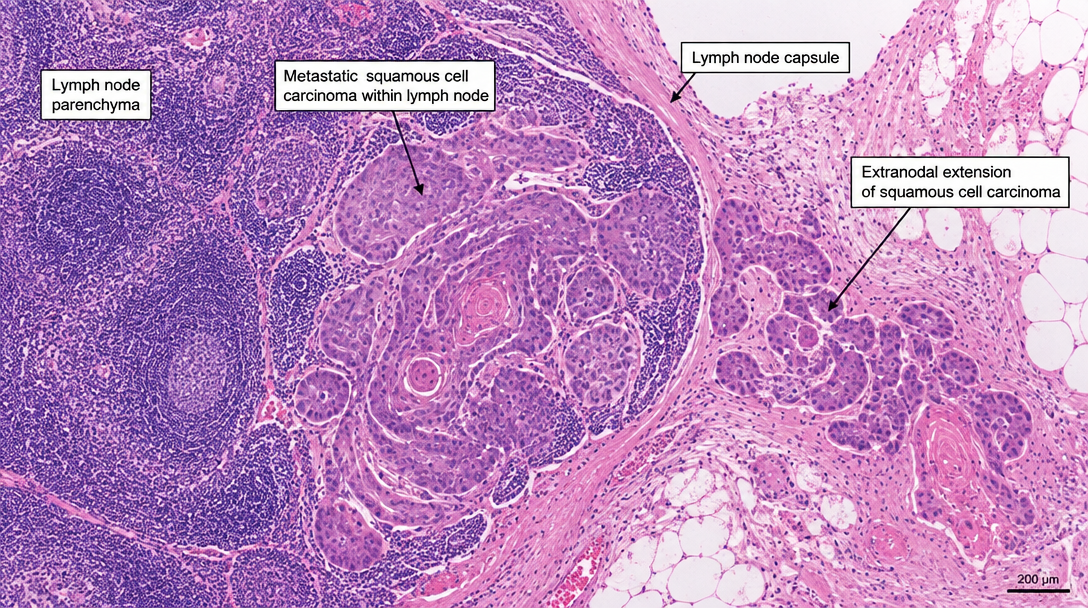
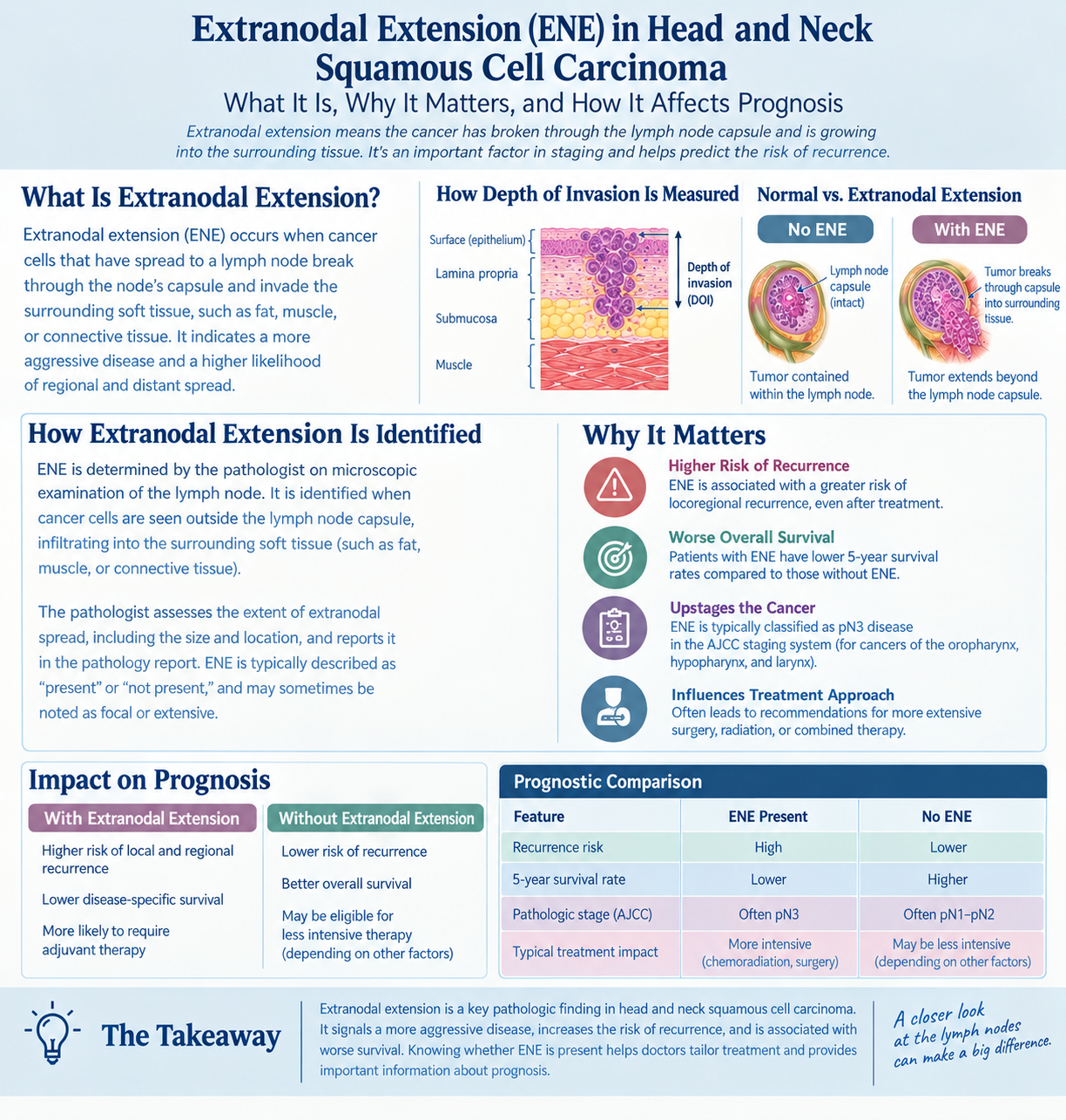
An extranodal extension lymph node finding starts with a lymph node metastasis. A lymph node is a small immune system filter, and cancer cells can travel there from a nearby tumor in the mouth, throat, voice box, salivary gland, skin, thyroid region, or other head and neck site. The lymph node normally has a thin outer covering called a capsule. When cancer grows through that capsule into the surrounding tissue, pathologists call it extranodal extension.
A simple analogy is a seed inside a small paper envelope. If the seed stays inside the envelope, the boundary is intact. If it breaks through the envelope and grows into the surrounding soil, that is similar to extracapsular spread. In a pathology report, the word “extranodal” means outside the node, and “extension” means the cancer has crossed that boundary.
Pathologists diagnose an extranodal extension lymph node by examining tissue under the microscope after a lymph node dissection or removal of a suspicious node. The pathologist looks for cancer cells at the edge of the lymph node and checks whether they extend into fat, muscle, connective tissue, or other nearby soft tissue. In head neck cancer, this detail matters because it helps the oncology team estimate the risk that cancer cells may be present beyond the tissue that was visibly removed. For patients trying to understand the structure of the report itself, What Is a Pathology Report? can help explain how these findings are organized.
Why Your Report Shows This Finding
A pathology report may show extranodal extension because the cancer cells in a lymph node metastasis have grown aggressively enough to cross the lymph node capsule. This can happen in several types of head neck cancer, including squamous cell carcinoma of the oral cavity, oropharynx, hypopharynx, and larynx, as well as selected skin cancers and salivary gland cancers. The finding is not something a patient caused. It reflects the biology and growth pattern of the tumor in that lymph node.
Under the microscope, pathologists look for a transition from cancer confined inside the lymph node to cancer infiltrating the surrounding tissue. Sometimes the capsule is easy to see, and the diagnosis of extracapsular spread is straightforward. Other times, the lymph node is almost completely replaced by tumor, the capsule is distorted, and the assessment requires careful judgment. The pathologist may also measure how far the cancer extends outside the node, although reporting practices can vary by tumor type and institution.
Other parts of the report help place an extranodal extension lymph node finding into context. These include the number of positive lymph nodes, the size of the largest lymph node metastasis, the primary tumor size, tumor depth, lymphovascular invasion, perineural invasion, and surgical margins. Surgical margins describe whether cancer is present at or near the edge of the removed tissue. Cancer staging uses these features together, rather than relying on one line of the report alone.
How Serious Is Extranodal Extension Lymph Node Involvement?
Extranodal extension lymph node involvement is usually considered a higher-risk finding in head neck cancer. It suggests that the cancer did not simply enter a lymph node and remain neatly contained there. Instead, it crossed a natural boundary and entered nearby tissue, which can raise concern for microscopic cancer cells in the regional area. This is why the finding often influences cancer staging and postoperative treatment recommendations.
The seriousness depends on the full clinical picture. A tiny focus of extranodal extension in one lymph node may carry a different level of risk than bulky disease involving multiple nodes with obvious soft tissue invasion. The type of primary tumor also matters, including whether an oropharyngeal squamous cell carcinoma is HPV-associated. Surgical margins, tumor location, imaging findings, and the patient’s overall health all help determine how much weight the team gives to the extranodal extension lymph node result.
In many head neck cancer treatment plans, extranodal extension is one of the reasons doctors may recommend radiation therapy after surgery, sometimes with chemotherapy. Radiation therapy is used to treat the area where microscopic cancer cells may remain, even when no visible cancer is left behind. This does not mean the surgery failed; it means the pathology report identified a risk factor that can guide additional treatment. Patients who want a more systematic way to read the full report may find Understanding Your Pathology Report: How to Read It with Confidence helpful before an oncology appointment.
What Happens Next: Treatment And Monitoring
After an extranodal extension lymph node diagnosis, the case is often reviewed by a multidisciplinary team. This may include head and neck surgeons, radiation oncologists, medical oncologists, radiologists, pathologists, dentists, speech and swallowing specialists, and nutrition experts. The team reviews the primary tumor, lymph node metastasis, surgical margins, imaging studies, and cancer staging together. The goal is to match the intensity of treatment to the actual risk shown by the tissue and imaging findings.
For many patients, the next step is a discussion about radiation therapy. In some cases, chemotherapy may be added to radiation therapy, especially when the risk of recurrence is considered high. The recommendation depends on the cancer type, the patient’s recovery from surgery, kidney function, hearing status, nerve function, swallowing ability, and other medical factors. Treatment decisions should be individualized rather than based on one phrase alone.
Monitoring after treatment usually includes physical examinations, imaging when appropriate, attention to swallowing and speech, dental care, and symptom review. The team watches for signs of recurrence and also manages treatment effects such as dry mouth, swallowing difficulty, neck stiffness, or fatigue. A pathology report with extranodal extension gives the team a map of where risk was highest. That map can help direct follow-up in a careful, targeted way.
Questions To Ask Your Doctor Or Pathologist
- How many lymph nodes were positive for cancer?
- Was extranodal extension described as microscopic, limited, extensive, or gross?
- What was the size of the largest lymph node metastasis?
- Were the surgical margins negative, close, or positive?
- How does this finding change cancer staging in this specific head neck cancer?
- Does this result mean radiation therapy or chemotherapy is recommended?
- Would a second pathology review be useful before final treatment planning?
These questions can help transform a difficult report into a focused conversation. Patients often hear several specialties discuss the same finding from different angles, and that can be confusing unless the pathology details are clear. A pathologist’s role is to define what was seen in the tissue as accurately as possible, so the oncology team can make informed treatment recommendations.
A second pathology opinion may be reasonable when the diagnosis is unexpected, the report language is unclear, the cancer type is unusual, or treatment would change based on the degree of extranodal extension. This is especially true in head neck cancer, where small differences in interpretation can affect cancer staging or postoperative treatment. For patients considering review, When Should You Get a Second Pathology Opinion? and How to Get a Second Opinion on Your Pathology Diagnosis explain the process in practical terms.
Frequently Asked Questions
What does extranodal extension in a lymph node mean?
It means cancer spread to a lymph node and then grew through the lymph node capsule into nearby tissue. An extranodal extension lymph node result is different from cancer that stays confined inside the node. In head neck cancer, this finding often increases concern for regional microscopic disease. The final meaning depends on the full pathology report, imaging, and clinical examination.
Is extranodal extension the same as extracapsular spread?
Yes, these terms are closely related and are often used to describe the same process. Extracapsular spread means cancer has moved beyond the capsule, or outer covering, of a lymph node. Many modern pathology reports use “extranodal extension” instead. The wording may vary, but the clinical concern is similar.
Does extranodal extension lymph node mean cancer is stage 4?
An extranodal extension lymph node finding can affect cancer staging, but it does not automatically mean the same stage for every patient. Staging rules differ by tumor site, HPV status in certain oropharyngeal cancers, and other factors. The number and size of involved lymph nodes also matter. The oncology team should explain how the finding changes the stage in the specific case.
Can extranodal extension be seen on a scan?
Sometimes imaging suggests extranodal extension, especially when a lymph node has irregular borders, invasion into nearby structures, or matted nodes. However, the most definite diagnosis usually comes from microscopic examination in the pathology report. A scan and the microscope answer related but different questions. When both suggest extracapsular spread, the finding may carry more confidence.
Can treatment still work if extranodal extension is present?
Yes, treatment can still be effective when extranodal extension is present. The finding often prompts doctors to recommend additional therapy, such as radiation therapy with or without chemotherapy, to reduce recurrence risk. An extranodal extension lymph node result is a risk factor, not a prediction that treatment will fail. The best next step is a clear discussion of the full treatment plan and its goals.
An extranodal extension lymph node finding is serious, but it is also actionable information. It helps the care team understand where the cancer has been and how aggressively the regional area may need to be treated. Honest Pathology consultations can help patients and families clarify the wording of a report, prepare better questions, and approach treatment discussions with more confidence.
References:
National Cancer Institute — Pathology Reports
National Cancer Institute — Definition of Lymph Node
National Cancer Institute — Head and Neck Cancers