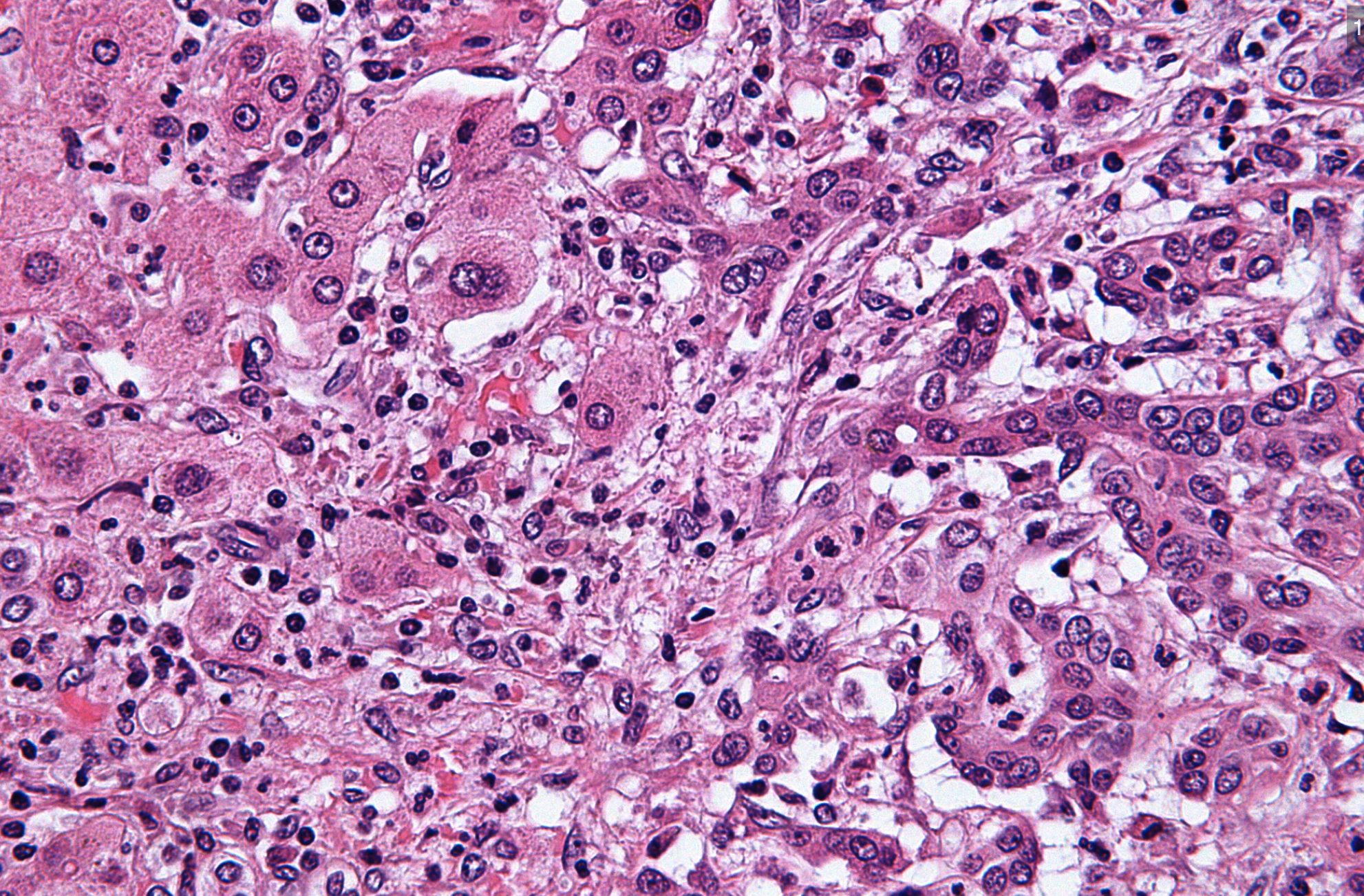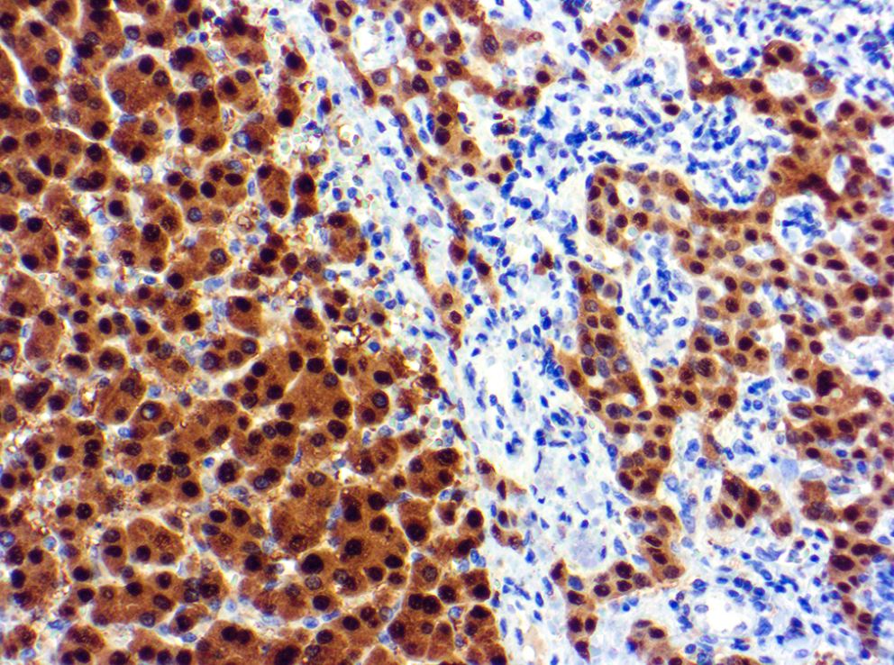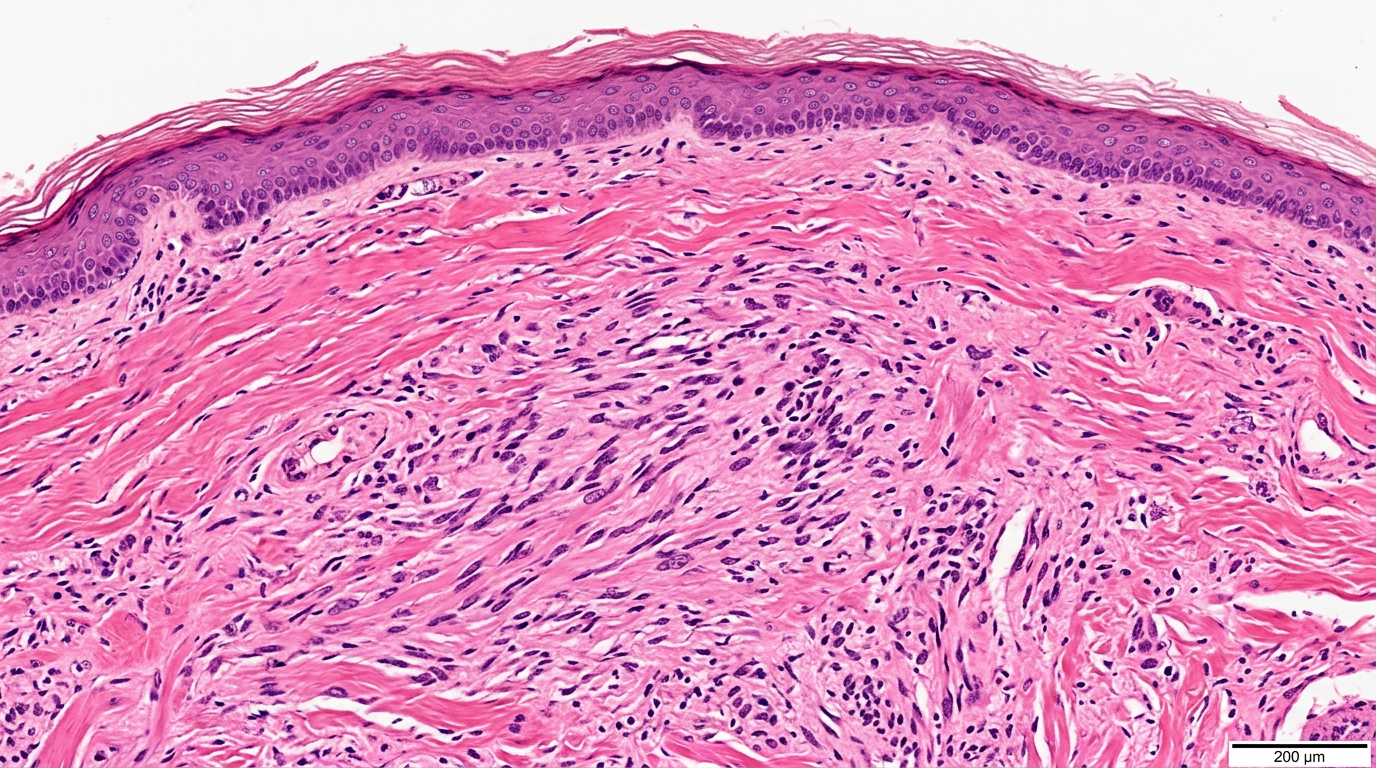
A melanoma report can feel frightening. desmoplastic melanoma diagnosis means a specific melanoma pattern; learn what to ask next.
desmoplastic melanoma diagnosis means the pathologist found a form of melanoma in which cancer cells grow within dense scar-like tissue, often in the skin of the head, neck, or sun-damaged areas. This diagnosis is made under the microscope, usually after a biopsy or excision, and it often requires careful review because it can look deceptively subtle. A melanoma pathology report may include details such as Breslow thickness, surgical margins, and perineural invasion because these features help guide treatment planning.
Hearing the word melanoma can make time feel like it stops. The phrase desmoplastic melanoma diagnosis may sound even more confusing because it is not a term most patients have heard before. Clarity can lower fear: The report is not just a label, but a map of what was found, how deeply it grew, and what decisions may come next. Patients deserve explanations that are precise, calm, and honest.
Desmoplastic Melanoma Diagnosis — What It Actually Means
A desmoplastic melanoma diagnosis describes a subtype of melanoma with a fibrous, firm, scar-like background. “Desmoplastic” means that the tumor stimulates dense collagen, the same type of structural material the body uses in scars. Under the microscope, the melanoma cells may be stretched out and thin, rather than forming the more obvious nests seen in many other melanomas. This is why spindle cell melanoma is a term that may appear in or near the report.
In practical terms, the pathologist is saying that the skin biopsy results show melanoma, but with a growth pattern that can be easy to underestimate clinically. Desmoplastic melanoma may look like a scar, a firm bump, a pale patch, or a slowly changing area rather than a classic dark mole. A melanoma pathology report usually pulls together the diagnosis, tumor depth, margin status, and other risk features. For patients trying to understand the document itself, a pathology report is best viewed as the medical translation of what the tissue shows.
One useful analogy is a weed growing through thick roots and soil. The visible surface may not fully show how the tumor extends through the deeper dermis. Pathologists examine thin stained sections to determine whether the tumor is confined, deeply invasive, close to an edge, or tracking near nerves. That is why Breslow thickness, surgical margins, and perineural invasion can strongly influence what the care team recommends.
Why Your Report Shows This Finding
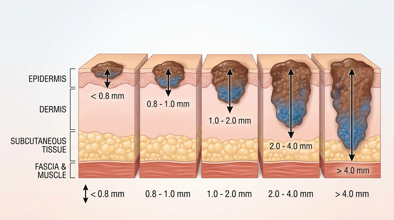
Desmoplastic melanoma is usually diagnosed because the biopsy shows atypical melanocytes growing as spindled cells within dense collagen. These cells may be less pigmented than typical melanoma cells, so the diagnosis may rely on special stains called immunohistochemistry. Markers such as SOX10 and S100 often help confirm spindle cell melanoma when routine microscope features are not enough. The melanoma pathology report may mention these tests if the tumor was difficult to classify.
Several factors make this tumor different from more familiar melanoma patterns. It often develops on chronically sun-damaged skin, especially in older adults, although it can occur in other settings. Nerve involvement is a recognized feature, and the report may use the term perineural invasion when melanoma cells are seen around or along a nerve. Perineural invasion matters because it can affect local recurrence risk and may influence surgery or radiation discussions.
The report may also describe Breslow thickness, ulceration, mitotic rate, lymphovascular invasion, and surgical margins. Breslow thickness measures how deep the melanoma extends from the skin surface and is one of the most important staging features. Surgical margins describe whether melanoma is present at, close to, or away from the cut edge of the specimen. Clear skin biopsy results do not always mean the first biopsy removed everything, because a biopsy may sample only part of the lesion.
How Serious Is a Desmoplastic Melanoma Diagnosis?
A desmoplastic melanoma diagnosis is serious because it is melanoma, but seriousness depends on the exact features in the report. Some cases are found early and can be treated effectively with complete surgical removal. Other cases are thicker, involve nerves, or extend close to surgical margins, which can require wider excision and careful follow-up. The most useful question is not simply whether it is serious, but which risk features are present.
Breslow thickness is central to that risk assessment. A thin melanoma generally carries a better outlook than a thick melanoma, although desmoplastic melanoma has special behavior that clinicians consider separately. Perineural invasion can increase concern for local recurrence, especially when there is extensive nerve involvement. A melanoma pathology report should be read with attention to both the diagnosis line and the microscopic details beneath it.
Another key issue is whether the tumor is pure desmoplastic melanoma or mixed desmoplastic melanoma. Pure desmoplastic melanoma is made almost entirely of the desmoplastic pattern, while mixed tumors also contain conventional melanoma areas. This distinction may affect lymph node risk and treatment planning. If the desmoplastic melanoma diagnosis was made on a small biopsy, a larger excision may provide more complete information about surgical margins, spindle cell melanoma pattern, and true depth.
What Happens Next: Treatment and Monitoring
After a desmoplastic melanoma diagnosis, treatment usually begins with ensuring complete removal of the tumor. The surgeon may recommend a wide local excision, which removes additional normal-looking skin around the melanoma to create a safety margin. The needed width depends on factors such as Breslow thickness and anatomic location. Surgical margins from the final excision help show whether the tumor appears fully removed.
The care team may also discuss sentinel lymph node biopsy, imaging, radiation therapy, or medical oncology referral depending on the report. Desmoplastic melanoma can have a lower lymph node spread rate in some pure cases, but decisions are individualized. Perineural invasion, deep growth, positive margins, and recurrence history may raise the need for additional treatment discussions. Nerve involvement is one reason radiation oncology may sometimes be considered, particularly in head and neck tumors.
Follow-up typically includes skin examinations, lymph node checks, and attention to the scar or surgical site. Patients may be asked to report new firmness, numbness, tingling, pain, or changes near the excision area. Skin biopsy results from future suspicious areas should be interpreted in the context of the prior melanoma history. For help reading report sections such as diagnosis, margins, and microscopic description, understanding a pathology report can make the next appointment more focused.
Questions to Ask Your Doctor or Pathologist
- Is this a pure or mixed desmoplastic melanoma diagnosis?
- What is the Breslow thickness, and how does it affect stage?
- Are the surgical margins clear, close, or involved by melanoma?
- Does the report show perineural invasion or other nerve involvement?
- Was immunohistochemistry used to confirm spindle cell melanoma?
- Is a sentinel lymph node biopsy recommended in this specific case?
- Should the slides be reviewed by a dermatopathologist or melanoma specialist?
These questions can turn a frightening appointment into a structured conversation. A melanoma pathology report contains many small details, and each detail may change the next step. If the original skin biopsy results are limited, the final excision may add essential information about depth and surgical margins.
A second review can be valuable when the diagnosis is rare, subtle, or treatment-changing. Desmoplastic melanoma can overlap microscopically with scars, nerve sheath tumors, and other spindle cell tumors, so expert confirmation may be appropriate. Patients considering this option may find guidance on when to get a second pathology opinion helpful before requesting slide review.
Frequently Asked Questions
Is desmoplastic melanoma cancer?
Yes. Desmoplastic melanoma is a type of melanoma, which is a cancer of melanocytes. A desmoplastic melanoma diagnosis means the cancer has a scar-like, fibrous growth pattern under the microscope. The behavior depends on depth, margins, perineural invasion, and whether the tumor is pure or mixed.
Can desmoplastic melanoma be mistaken for a scar?
Yes, both clinically and microscopically it can resemble scar tissue. The tumor may be pale, firm, or subtle, and spindle cell melanoma can blend into dense collagen. Pathologists often use immunohistochemistry to separate melanoma from scar and other spindle cell tumors. This is one reason a melanoma pathology report may be more detailed than expected.
Does perineural invasion mean melanoma has spread?
Perineural invasion means melanoma cells are growing around or along a nerve in the tissue examined. It does not automatically mean distant spread to organs. It may increase concern for local recurrence and can influence surgery, radiation, and follow-up decisions. Nerve involvement should be discussed with the treating team in the context of the whole report.
What is the best treatment for desmoplastic melanoma?
The main treatment is usually complete surgical removal with appropriate surgical margins. Additional treatment depends on Breslow thickness, margin status, perineural invasion, tumor location, and stage. Some patients may need sentinel lymph node biopsy, radiation therapy, immunotherapy, or imaging. A desmoplastic melanoma diagnosis should be managed by a team familiar with melanoma care.
Should I get a second opinion for desmoplastic melanoma diagnosis?
A second opinion can be reasonable because this is an uncommon and sometimes subtle melanoma subtype. A desmoplastic melanoma diagnosis may affect surgery, lymph node evaluation, radiation decisions, and follow-up intensity. The original slides can often be reviewed without repeating the biopsy. Practical steps are outlined in how to get a second opinion on a pathology diagnosis.
A desmoplastic melanoma diagnosis can feel overwhelming, but the report contains clues that help the care team choose the safest next step. The most helpful path forward is to clarify the depth, margins, nerve findings, and whether expert slide review is needed. Honest Pathology consultations can help patients and families understand the pathology language before major treatment decisions.
References:
National Cancer Institute — Pathology Reports Fact Sheet
National Cancer Institute — Melanoma Definition
NCBI Bookshelf — Clinical Medical References