Worried by confusing biopsy wording? desmoid fibromatosis sarcoma differences explain risk, diagnosis, and what to ask next.
desmoid fibromatosis sarcoma differences matter because desmoid-type fibromatosis can look and behave aggressively in one area, but it is not the same diagnosis as a true sarcoma. A pathology report may use terms such as “fibromatosis,” “locally aggressive,” “spindle cell lesion,” or “rule out sarcoma,” which can feel frightening when read without context. Pathologists separate these diagnoses by studying the cells, growth pattern, stains, and sometimes molecular findings.
Fear is a very normal reaction when a report mentions a soft tissue tumor and sarcoma in the same clinical conversation. The key point is that pathology language often describes a differential diagnosis, not a final verdict. A careful review can turn a vague phrase into a clearer answer, and that clarity can change what treatment is considered safest.
Desmoid-type fibromatosis sits in an unusual middle ground: It can grow into nearby tissue and come back locally, but it does not spread to distant organs like sarcoma. That difference is the reason expert interpretation matters so much.
Desmoid Fibromatosis Sarcoma — What It Actually Means
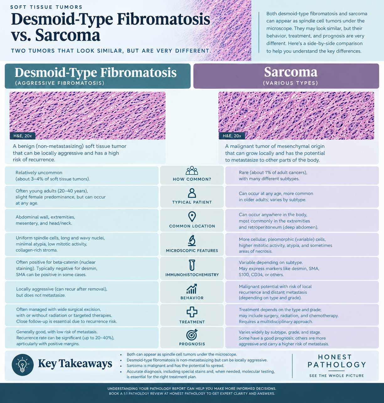
The phrase desmoid fibromatosis sarcoma usually reflects confusion between two different categories of soft tissue disease. Desmoid-type fibromatosis is a clonal fibroblastic tumor that grows in connective tissue, while sarcoma is a malignant soft tissue tumor with the potential to metastasize. In plain language, desmoid-type fibromatosis may act like an invasive vine in a garden, wrapping into nearby structures, but it does not usually send seeds to distant sites. Sarcoma is different because it can invade locally and, depending on type and grade, spread through the bloodstream.
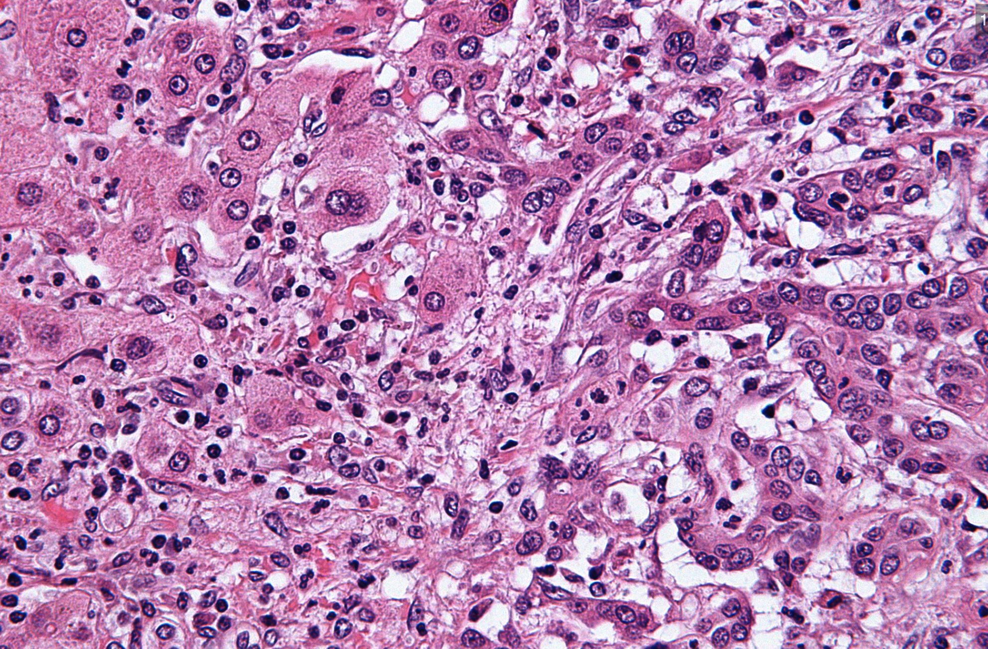
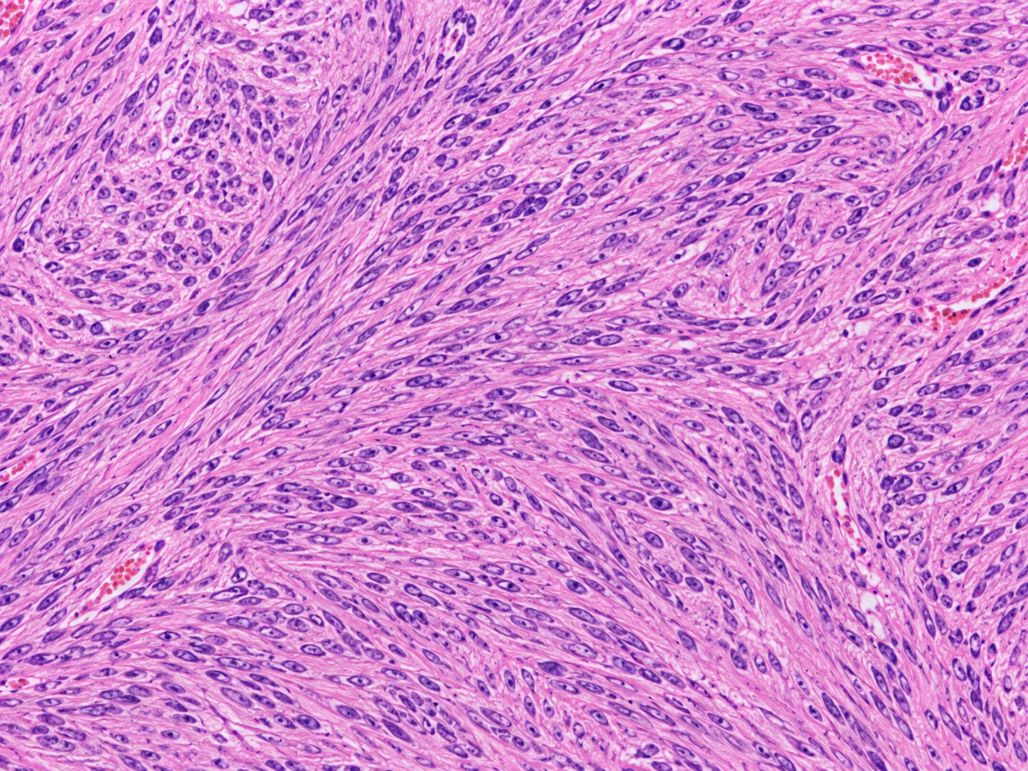
Under the microscope, pathologists look for spindle cells arranged in long sweeping bundles within collagen-rich tissue. These spindle cells can appear bland in desmoid-type fibromatosis, meaning they do not show the severe nuclear abnormalities expected in many sarcomas. A pathology report may describe low cellular atypia, rare mitoses, and infiltrative edges. The words can sound alarming, but each term helps separate a locally aggressive process from a high-grade cancer.
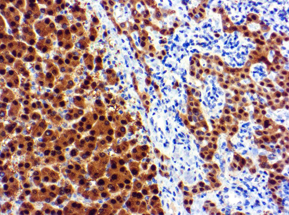
The beta catenin stain is often used because many desmoid tumors show nuclear beta-catenin staining. This beta catenin stain is not interpreted alone, but it can support the diagnosis when the microscopic pattern fits. A pathologist also considers the biopsy site, imaging findings, and whether the sample is large enough to represent the mass. For patients trying to decode the first document, what a pathology report contains can make the wording less intimidating.
Why Your Report Mentions Both Diagnoses
A report may mention both desmoid-type fibromatosis and sarcoma because small biopsies sometimes sample only part of a lesion. Many soft tissue tumors are made of spindle cells, so the first job is to identify whether the cells look bland, atypical, inflammatory, reactive, or frankly malignant. A desmoid fibromatosis sarcoma concern often appears when the tumor is deep, large, painful, recurrent, or located near critical structures. Those clinical features can overlap, even when the final diagnoses are biologically different.
Pathologists use routine hematoxylin and eosin slides first, then add immunohistochemistry when needed. The beta catenin stain can show nuclear staining in desmoid-type fibromatosis, while other stains may help exclude nerve sheath tumor, smooth muscle tumor, solitary fibrous tumor, or specific sarcoma types. Molecular testing may identify a CTNNB1 mutation in sporadic desmoid-type fibromatosis. In some cases, APC-associated disease is considered, especially when the tumor occurs in the abdomen or in the setting of familial adenomatous polyposis.
The phrase “cannot exclude sarcoma” does not always mean that sarcoma is likely. It may mean the biopsy is limited, the tissue is crushed, the lesion is unusually cellular, or the imaging appearance is more aggressive than expected. Surgical margins may also be mentioned if the lesion has already been removed, because desmoid-type fibromatosis often extends into surrounding tissue in a finger-like way. A clear understanding of the pathology report and imaging together is usually stronger than either one alone, and learning how to read a pathology report with confidence can help families prepare better questions.
How Serious Is Desmoid Fibromatosis Sarcoma Confusion?
Desmoid fibromatosis sarcoma confusion is serious because the treatment paths can be very different. A true sarcoma may require staging, tumor grading, surgery, radiation, chemotherapy, targeted therapy, or referral to a sarcoma oncology team. Desmoid-type fibromatosis may be managed with active surveillance, medication, local therapy, surgery in selected cases, or multidisciplinary review. Treating desmoid-type fibromatosis like a high-grade sarcoma can expose a patient to unnecessary harm, while missing a sarcoma can delay needed cancer care.
The most reassuring distinction is that desmoid-type fibromatosis does not metastasize. That means it does not behave like a classic malignant sarcoma that spreads to the lungs or other organs. However, “not metastatic” does not mean “not important.” A desmoid tumor can cause pain, bowel obstruction, nerve compression, limited movement, or repeated local recurrence, depending on where it grows.
The seriousness depends on location, symptoms, growth rate, and diagnostic certainty. A small abdominal wall desmoid may be watched carefully, while a deep tumor near major nerves or organs may need more urgent multidisciplinary planning. Surgical margins can be difficult because the tumor may blend into normal fascia or muscle, and wider surgery is not always the best answer. When the report uses uncertain language, a second opinion from a pathologist experienced in soft tissue tumor diagnosis is often reasonable.
Desmoid fibromatosis sarcoma wording can also affect emotions, insurance authorization, and treatment referrals. Some patients hear the word sarcoma before the final diagnosis is complete, then carry that fear even after the pathology is clarified. A second opinion can confirm the diagnosis, recommend additional stains, or suggest molecular testing for a CTNNB1 mutation. The goal is not to create doubt for its own sake; the goal is to make sure the label matches the biology.
What Happens Next: Treatment And Monitoring
After a diagnosis is clarified, the next step is usually a treatment discussion that includes pathology, imaging, symptoms, and patient goals. Desmoid-type fibromatosis is often discussed in a multidisciplinary setting because surgery is not automatically the first choice. Many centers now consider active surveillance for selected patients, especially when the tumor is stable or symptoms are mild. This approach can feel surprising, but it reflects the unpredictable natural history of some desmoid tumors, including periods of no growth or even regression.
If the lesion is growing or causing symptoms, treatment may include nonsteroidal anti-inflammatory drugs, hormonal approaches in selected situations, tyrosine kinase inhibitors, newer targeted agents, radiation therapy, ablation, or surgery. The pathology report still matters because treatment decisions depend on confidence in the diagnosis. A soft tissue tumor with atypical features may require additional review before a conservative plan is chosen. The beta catenin stain, other immunostains, and molecular findings all help place the tumor in the correct category.
Monitoring commonly includes physical examination and MRI or CT imaging at intervals chosen by the treating team. Surgical margins may be followed closely when an excision has been performed, because local recurrence is a known risk. Pathology review can also be useful if the tumor recurs, grows faster than expected, or shows features that do not match the original diagnosis. Patients considering expert review can learn more about when a second pathology opinion may help.
Questions To Ask Your Doctor Or Pathologist
- Does the pathology report give a final diagnosis of desmoid-type fibromatosis, or is sarcoma still in the differential diagnosis?
- Were the cells described as bland spindle cells, atypical spindle cells, or high-grade malignant cells?
- Was a beta catenin stain performed, and did it show nuclear staining?
- Was molecular testing for a CTNNB1 mutation considered or performed?
- Do the imaging findings match the pathology diagnosis of this soft tissue tumor?
- If surgery was done, what do the surgical margins show, and how does that affect recurrence risk?
- Would a second opinion from a soft tissue pathologist be useful before treatment begins?
These questions are not meant to challenge the treating team. They help translate a complex pathology report into decisions about surveillance, surgery, radiation, or systemic therapy. Desmoid fibromatosis sarcoma wording should be clarified before major treatment whenever possible.
A second opinion is especially helpful when the biopsy is small, the tumor is deep, the report uses uncertain language, or treatment would be intensive. The process usually involves sending slides, blocks, reports, and imaging information to another pathology service. Patients who need a practical roadmap can review how to get a second opinion on a pathology diagnosis.
Frequently Asked Questions
Is desmoid-type fibromatosis a cancer?
Desmoid-type fibromatosis is usually described as an intermediate or locally aggressive tumor rather than a metastatic cancer. It can grow into nearby tissue and recur in the same area, but it does not spread to distant organs like most sarcomas can. This is why desmoid fibromatosis sarcoma wording needs careful explanation. The diagnosis can still be clinically serious depending on location and symptoms.
Can desmoid fibromatosis be mistaken for sarcoma?
Yes, desmoid-type fibromatosis can sometimes be confused with sarcoma, especially on a small biopsy. Both can be spindle cell lesions, and both may occur in deep soft tissue. The beta catenin stain, microscopic features, imaging correlation, and sometimes molecular testing help separate them. A second opinion may be appropriate when the pathology report is uncertain.
What does beta-catenin positive mean in desmoid tumor?
Beta-catenin positive usually means that the beta catenin stain shows staining in the nuclei of tumor cells. In the right microscopic setting, this supports desmoid-type fibromatosis. It is not a perfect test by itself, so pathologists interpret it with the tissue pattern and clinical information. Molecular testing for a CTNNB1 mutation can provide additional support in selected cases.
Does desmoid fibromatosis need surgery?
Not always. Many patients with desmoid-type fibromatosis are considered for active surveillance first, especially when the tumor is stable and symptoms are manageable. Surgery may be considered when the tumor causes significant problems or when other options are not suitable. Surgical margins matter, but aggressive surgery is not always the safest or most effective plan.
Should I get a second opinion for desmoid fibromatosis sarcoma?
A second opinion can be very helpful when the diagnosis includes desmoid fibromatosis sarcoma uncertainty, limited biopsy tissue, unusual staining, or major treatment decisions. Soft tissue pathology is a specialized area, and expert review may confirm the diagnosis or recommend additional tests. The review can also clarify whether the report supports desmoid-type fibromatosis, sarcoma, or another soft tissue tumor. Clear diagnosis helps the treating team choose a plan with more confidence.
A confusing soft tissue diagnosis can feel overwhelming, but the words in a report are pieces of evidence, not a life sentence. Desmoid-type fibromatosis and sarcoma are separated by careful pathology review, clinical correlation, and sometimes molecular testing. Honest Pathology consultations can help patients and families understand the report, organize questions, and decide whether expert review is appropriate.
References:
National Cancer Institute — Pathology Reports
National Cancer Institute — Sarcoma Definition
NCBI Bookshelf — Medical Reference Texts