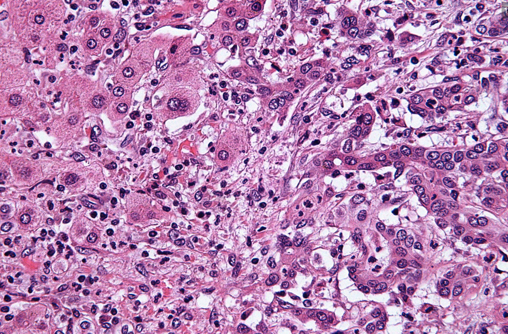
A confusing biopsy result can feel frightening. atypical lymphoid proliferation means uncertainty, not always cancer. Learn what to ask next.
atypical lymphoid proliferation means that a biopsy shows lymphoid immune cells with unusual features, but the findings are not automatically the same as lymphoma. Pathologists use this phrase when the tissue raises concern, yet the available sample, cell pattern, or test results do not allow a clean benign-versus-malignant answer. It is a diagnostic signal that more context, more testing, or sometimes another biopsy may be needed.
Receiving this wording in a pathology report can be deeply unsettling because it sounds technical and unfinished. Many patients read it as a hidden cancer diagnosis, but that is not always correct. The phrase often sits in the gray zone between reactive lymphocytes, which can appear during infection or inflammation, and a true lymphoma diagnosis. Clear next steps can turn that uncertainty into a plan.
A biopsy report is not just a label; it is a record of what the pathologist saw under the microscope, what tests were performed, and what questions remain. A helpful starting point is learning how a pathology report is structured, because wording such as atypical, suspicious, or cannot exclude has specific meaning in pathology.
Atypical Lymphoid Proliferation — What It Actually Means
Atypical Lymphoid Proliferation describes an abnormal-looking collection of lymphoid cells, the immune cells that normally live in lymph nodes, tonsils, spleen, bone marrow, skin, stomach, and many other tissues. The word atypical means the cells or their pattern do not look entirely usual. The word proliferation means there are more of these cells than expected or they are arranged in a pattern that draws attention. This can happen in a lymph node biopsy, a skin biopsy, a stomach biopsy, or a biopsy from another site.
A useful analogy is a smoke detector. Smoke may come from a fire, but it may also come from burnt toast, steam, or dust. In the same way, atypical lymphoid proliferation tells the medical team that the biopsy has triggered concern, but it does not always prove lymphoma. The pathologist then checks the architecture of the tissue, the size and shape of the cells, and whether the cells behave like reactive lymphocytes or like a clonal population. The final meaning depends on the full pathology report, not the phrase alone.
In many cases, the pathologist adds a comment explaining whether the finding is favored to be reactive, suspicious for lymphoma, or limited by the sample size. A small needle sample may not show enough tissue architecture to make a confident lymphoma diagnosis. A larger excisional lymph node biopsy may answer the question more clearly because it preserves the overall structure. When the report mentions flow cytometry or immunohistochemistry stains, those tests are being used to sort the immune cells into more specific groups.
Why The Report Shows This Finding

A pathology report may show atypical lymphoid proliferation for several reasons. Some causes are benign, including viral infections, autoimmune disease, drug reactions, immune stimulation, or inflammation near a tumor or wound. In these situations, reactive lymphocytes can enlarge, multiply, and look worrisome even though they are part of a normal immune response. The microscopic pattern may overlap with early lymphoma, which is why the wording can be cautious.
Other times, atypical lymphoid proliferation may be used when lymphoma is a real possibility but not fully proven. The pathologist may see a dense lymphoid infiltrate, abnormal follicles, large atypical cells, or an uneven mix of B cells and T cells. Immunohistochemistry stains help show which cell types are present and whether they are arranged normally. Flow cytometry can look for an abnormal immunophenotype, and it may identify a clonal population of B cells or T cells.
The biopsy site also matters. A lymph node biopsy is interpreted differently from a small skin biopsy or a stomach biopsy because each tissue has its own normal immune background. In the stomach, for example, chronic inflammation can produce lymphoid aggregates that mimic a low-grade lymphoma. In skin, some inflammatory rashes can contain many atypical lymphocytes. For that reason, the pathologist often combines microscopic findings with clinical information such as symptoms, imaging, blood counts, infection testing, and whether lymph nodes are enlarging over time.
How Serious Is Atypical Lymphoid Proliferation?
The seriousness of atypical lymphoid proliferation depends on what the report says around the phrase. If the comment states that the findings are favored reactive, and the patient has a recent infection or inflammatory condition, the chance of a lymphoma diagnosis may be lower. If the report says suspicious for lymphoma, cannot exclude lymphoma, or recommends additional tissue, the concern is higher. The phrase itself is not a stage, not a grade, and not a final cancer category.
Atypical lymphoid proliferation becomes more concerning when several features line up in the same direction. These features may include a persistent enlarged lymph node, abnormal imaging, unexplained fevers, night sweats, weight loss, abnormal blood counts, a clonal population on flow cytometry, or strongly abnormal immunohistochemistry stains. A lymph node biopsy that shows loss of normal architecture can also raise concern. Still, even in this setting, the medical team usually needs a precise lymphoma diagnosis before treatment decisions can be made.
There are also mild or uncertain scenarios. Sometimes a tiny biopsy captures only part of a reactive process, leaving the pathologist unable to be definitive. Sometimes prior steroids, antibiotics, or partial treatment change the appearance of the tissue. Sometimes flow cytometry is negative because the abnormal cells are too few, fragile, or not sampled. In those cases, atypical lymphoid proliferation is a careful statement of uncertainty rather than a final answer.
Patients often want to know whether this means cancer is being hidden from them. In pathology, uncertainty is usually stated plainly because accurate classification matters. A lymphoma diagnosis affects treatment, prognosis, and follow-up, so pathologists avoid overcalling lymphoma when the evidence is incomplete. At the same time, they avoid dismissing an abnormal pattern when more evaluation is needed.
What Happens Next: Treatment And Monitoring
The next step after atypical lymphoid proliferation is usually correlation, which means comparing the biopsy with the patient’s history, exam, imaging, blood tests, and prior pathology report results. The treating clinician may order complete blood counts, viral studies, autoimmune testing, imaging, or repeat examination of the affected area. If the concern is low, the plan may be observation with follow-up rather than immediate treatment. If the concern is higher, a hematologist, oncologist, dermatologist, gastroenterologist, or surgeon may become involved depending on the biopsy site.
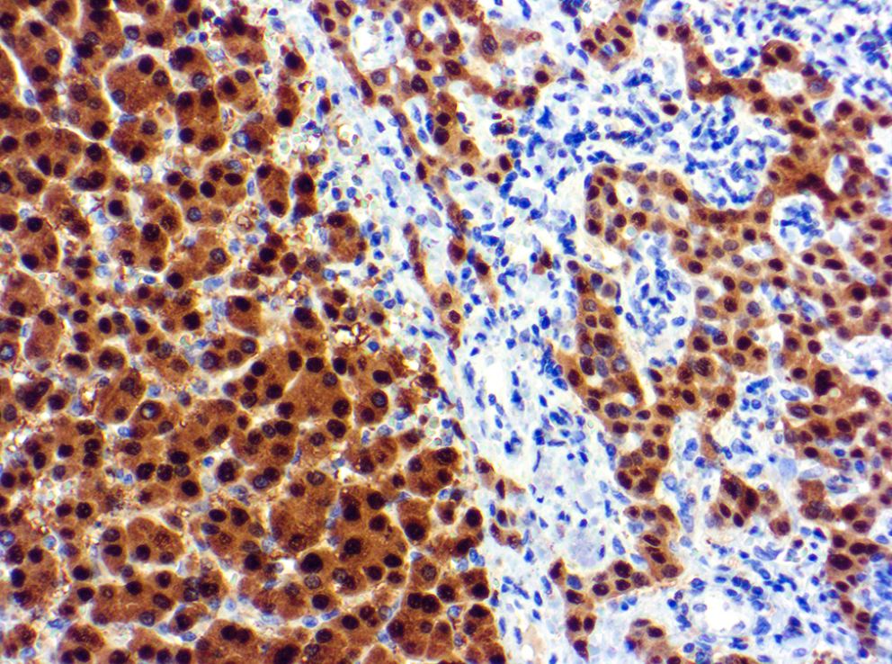
Additional pathology testing is common. Immunohistochemistry stains may be added to evaluate B-cell markers, T-cell markers, proliferation rate, and patterns that support or argue against lymphoma. Flow cytometry may be performed if fresh tissue was available, and molecular studies may look for gene rearrangements that suggest a clonal population. These tests do not replace the microscope; they add layers of evidence to the original tissue interpretation.
Sometimes the most useful next step is another biopsy. A repeat lymph node biopsy, especially an excisional biopsy that removes an entire node, may provide enough architecture for a definitive lymphoma diagnosis or reassurance. In other cases, the clinician may choose to repeat imaging after a short interval to see whether the lymph node shrinks, stays stable, or grows. Treatment is generally not started based only on the phrase atypical lymphoid proliferation unless the overall clinical and pathology evidence strongly supports a specific disease.
Questions To Ask Your Doctor Or Pathologist
- Does the pathology report favor a reactive process, lymphoma, or an uncertain gray-zone result?
- Was the biopsy sample large enough to evaluate tissue architecture, especially if this was a lymph node biopsy?
- Were immunohistochemistry stains performed, and what did they show about B cells and T cells?
- Was flow cytometry performed, and did it show a clonal population?
- Does the report recommend molecular testing, repeat biopsy, or clinical follow-up?
- Are symptoms, imaging findings, or blood counts increasing concern for a lymphoma diagnosis?
- Would a second pathology opinion from a hematopathologist be useful before treatment decisions?
These questions help move the conversation from fear to specifics. A patient does not need to master every marker or stain, but understanding whether the result is favored reactive, suspicious, or unresolved can make the next appointment more productive. The article on understanding a pathology report can also help patients identify the diagnosis, comment, and recommendation sections.
A second review can be especially helpful when the diagnosis is uncertain, the biopsy is small, or major treatment is being considered. Hematopathology is a subspecialty focused on lymph nodes, blood, bone marrow, and lymphoid diseases. Guidance on when to get a second pathology opinion and how to get a second opinion on a pathology diagnosis may be useful when the wording feels unresolved.
Frequently Asked Questions
Is atypical lymphoid proliferation cancer?
Atypical lymphoid proliferation is not automatically cancer. It means the biopsy shows lymphoid cells that look unusual or are arranged in a concerning pattern. Some cases are reactive lymphocytes from infection or inflammation, while others may represent early or partial lymphoma. The surrounding pathology report language and any added tests determine the level of concern.
Can atypical lymphoid proliferation turn into lymphoma?
Atypical lymphoid proliferation does not necessarily turn into lymphoma. In some cases, it is the first biopsy clue to an existing lymphoma that needs more tissue or testing to prove. In other cases, the finding resolves because it was related to infection, immune stimulation, or inflammation. Follow-up is meant to separate these possibilities safely.
Why would a biopsy be inconclusive for lymphoma?
A biopsy may be inconclusive when the tissue sample is very small, crushed, partially treated, or taken from an area that does not show the full process. Lymphoma diagnosis often depends on architecture, meaning how cells are organized across the tissue. A needle biopsy may miss the most abnormal area, while an excisional lymph node biopsy may show the pattern more clearly. Additional immunohistochemistry stains, flow cytometry, or molecular testing may be needed.
Should a second opinion be requested for atypical lymphoid proliferation?
A second opinion is reasonable when atypical lymphoid proliferation appears in a report and the next step could involve major treatment, repeat biopsy, or long-term surveillance. A hematopathologist may review the slides, stains, flow cytometry results, and clinical details. The review may confirm the original impression, refine the diagnosis, or recommend additional testing. A second opinion is not a sign of distrust; it is a quality step for a difficult diagnosis.
What tests help clarify atypical lymphoid proliferation?
Tests that may clarify atypical lymphoid proliferation include immunohistochemistry stains, flow cytometry, molecular clonality testing, blood tests, and imaging. Each test answers a different question about the lymphoid cells and their pattern. Flow cytometry can detect abnormal cell populations, while molecular testing may support the presence of a clonal population. The best test depends on the biopsy site, sample type, and level of suspicion in the pathology report.
A report that says atypical lymphoid proliferation can feel like being left halfway across a bridge. The safest next step is not panic, but clarification: the exact wording, the stains performed, whether flow cytometry was done, and whether more tissue or expert review is recommended. Honest Pathology consultations can help patients and families understand what the report is saying, what it is not saying, and which questions deserve priority at the next visit.
References:
National Cancer Institute — Pathology Reports Fact Sheet
National Cancer Institute — Lymphoma Definition
National Cancer Institute — Biopsy Definition