A brain tumor report can feel frightening. 1p/19q codeletion brain tumor results have specific meaning. Learn what to ask next.
1p/19q codeletion brain tumor means that testing found a specific combined loss of genetic material from chromosome 1 and chromosome 19 in the tumor cells. This result is most closely linked with oligodendroglioma, a type of diffuse glioma that also usually has an IDH mutation. In modern brain tumor pathology, this finding is not a small side note; it can help confirm the diagnosis, guide treatment planning, and give doctors useful information about expected behavior.
Seeing chromosome numbers and molecular terms on a report can make an already frightening diagnosis feel even more confusing. Many patients and families worry that “codeletion” means the tumor is automatically worse, but that is not usually how this result is interpreted. Pathologists use this result together with the microscope findings, imaging, tumor grading, and clinical details. A clear explanation can turn a technical phrase into a practical roadmap for the next appointment.
1p/19q Codeletion Brain Tumor — What It Actually Means
A 1p/19q codeletion brain tumor has lost two specific pieces of genetic material: The short arm of chromosome 1, called 1p, and the long arm of chromosome 19, called 19q. The word “codeletion” simply means both losses are present together. This is different from a single random genetic change and has a strong connection to oligodendroglioma diagnosis. In plain language, it is like finding a very specific fingerprint that helps identify the tumor family.
Pathologists do not diagnose a brain tumor from one molecular marker alone. The tissue is examined under the microscope, often with special stains, and then molecular testing helps refine the final classification. An IDH mutation is usually required along with whole-arm 1p and 19q codeletion for an oligodendroglioma diagnosis under current classification systems. This combined approach helps avoid older, less precise labels that were based mainly on appearance.
The phrase can sound alarming because it includes the word “deletion.” In this setting, however, the deletion is not something inherited from a parent or present throughout the body. It is usually a change found in the tumor cells themselves. For many patients, a 1p/19q codeletion brain tumor is associated with better response to certain treatments compared with similar diffuse gliomas that lack this pattern. For a broader foundation, patients may find it helpful to review what a pathology report is and how different sections fit together.
Why Your Report Shows This Finding
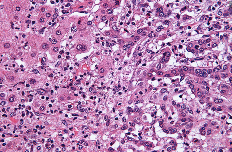
This result appears on a report because brain tumor pathology now depends on both appearance and molecular features. Under the microscope, some tumors show cells with rounded nuclei, delicate branching blood vessels, and other features that suggest oligodendroglioma. Those features raise the possibility, but they are not enough by themselves in many cases. Molecular testing is performed to look for IDH mutation and the characteristic loss of chromosome arms 1p and 19q.
Different laboratories may test for 1p/19q codeletion using methods such as fluorescence in situ hybridization, chromosomal microarray, next-generation sequencing, or other validated assays. The report may describe “whole-arm” loss because the key finding involves large chromosome arms, not just tiny fragments. This distinction matters because partial losses can occasionally mimic the pattern but may not carry the same diagnostic meaning. The pathology team interprets the technical result in the context of the tumor sample and the clinical picture.
Other results may appear nearby, including ATRX, p53, TERT promoter status, tumor grading features, and sometimes MGMT methylation. MGMT methylation is a different marker that may help predict response to temozolomide in certain gliomas, but it does not replace 1p/19q testing. Tumor grading is also separate from molecular classification; a tumor can be oligodendroglioma, IDH-mutant, and 1p/19q-codeleted, while still being assigned a grade based on mitotic activity, necrosis, microvascular proliferation, and other features. For patients trying to read multiple sections at once, understanding a pathology report can make the terminology less overwhelming.
How Serious Is a 1p/19q Codeletion Brain Tumor?
A 1p/19q codeletion brain tumor is serious because it is still a brain tumor, but the codeletion often carries more favorable information than many patients expect. In diffuse gliomas, the combination of IDH mutation and 1p/19q codeletion generally points toward oligodendroglioma, a tumor type that often grows more slowly and may respond well to radiation and chemotherapy. This does not mean the tumor is harmless or that treatment can be ignored. It means the diagnosis gives the care team more specific information than the word “glioma” alone.
The seriousness depends heavily on tumor grading, location, symptoms, imaging, patient age, performance status, and how much tumor can be safely removed. A lower-grade oligodendroglioma diagnosis may be monitored or treated differently from a higher-grade tumor with brisk cell division, necrosis, or microvascular proliferation. Brain tumor pathology helps identify these features, but the neurosurgeon and neuro-oncology team also consider whether the tumor affects speech, movement, seizures, or other critical functions. A 1p/19q codeletion brain tumor in a surgically difficult location may require a different plan than a similar tumor in a more accessible area.
Patients sometimes ask whether this result is “good news.” In context, the answer is often that it is a more favorable molecular finding within a difficult diagnosis. Molecular testing has shown that tumors with IDH mutation and 1p/19q codeletion often have a different clinical course than IDH-wildtype glioblastoma. Still, every case deserves careful review because small biopsy samples can miss high-grade areas, and older reports may use terminology that has changed. If the diagnosis, grade, or molecular testing result seems unclear, a second pathology opinion can be especially valuable; guidance is available on when to get a second pathology opinion.
What Happens Next: Treatment and Monitoring
After a 1p/19q codeletion brain tumor is reported, the next step is usually a combined discussion among neurosurgery, neuro-oncology, radiation oncology, radiology, and pathology. The care team reviews the surgery or biopsy results, MRI findings, tumor grading, and molecular testing. Treatment may include observation, additional surgery, radiation therapy, chemotherapy, or a combination of these approaches. The exact plan depends on the full clinical picture rather than the codeletion result alone.
Chemotherapy regimens such as PCV, which includes procarbazine, lomustine, and vincristine, have historically been important in treating 1p/19q-codeleted oligodendrogliomas. Temozolomide may also be discussed in some settings, and MGMT methylation may be part of that conversation. The treating team weighs benefits, side effects, age, symptoms, tumor size, and timing. The presence of 1p/19q codeletion brain tumor results can help doctors explain why certain treatments are being considered.
Monitoring usually involves regular MRI scans and neurologic follow-up. The interval between scans may be shorter soon after diagnosis or treatment and longer during stable periods. Seizure control, steroid use, cognitive changes, and rehabilitation needs may also be reviewed. Brain tumor pathology results should remain available because future decisions often depend on the original oligodendroglioma diagnosis, IDH mutation status, molecular testing method, and tumor grading.
Questions to Ask Your Doctor or Pathologist
- Does the report confirm an oligodendroglioma diagnosis, or is the diagnosis still uncertain?
- Was an IDH mutation found, and which testing method was used?
- Does the report show whole-arm 1p and 19q codeletion rather than a partial deletion?
- What is the tumor grading result, and were any high-grade features present?
- Was MGMT methylation tested, and does it affect treatment planning in this case?
- Was the sample large enough to confidently represent the tumor?
- Would a second pathology review or additional molecular testing change management?
These questions help translate a technical report into a practical conversation. Patients do not need to memorize every chromosome detail before an appointment, but having the key questions written down can reduce the feeling of being rushed or lost.
A second review is not a sign of distrust; it is a normal part of careful cancer care when the diagnosis has major treatment implications. For patients considering that step, practical instructions are available in how to get a second opinion on a pathology diagnosis.
Frequently Asked Questions
Is 1p/19q codeletion brain tumor good or bad?
A 1p/19q codeletion brain tumor is still a serious diagnosis because it involves a tumor in the brain. However, the codeletion often points toward oligodendroglioma, which may behave more favorably than some other diffuse gliomas. The result can also help guide treatment choices. The grade, MRI findings, symptoms, and surgical results still matter greatly.
Does 1p/19q codeletion mean oligodendroglioma?
1p/19q codeletion strongly supports an oligodendroglioma diagnosis when it occurs with an IDH mutation and the right pathologic features. Current classification systems rely on this molecular combination rather than microscope appearance alone. If IDH mutation is absent, the diagnosis should be reviewed carefully. Molecular testing helps make the diagnosis more precise.
Can a 1p/19q codeletion brain tumor be cured?
A 1p/19q codeletion brain tumor can often be treated effectively, sometimes for long periods, but the word “cure” depends on the individual situation. Diffuse gliomas can send tumor cells into surrounding brain tissue, which makes complete removal challenging. Some patients have years of stable disease after surgery and treatment. The treating neuro-oncology team can explain the expected course based on grade and imaging.
Is 1p/19q codeletion inherited?
In most cases, 1p/19q codeletion is a change found only in the tumor cells. It is not usually inherited from a parent and does not usually mean family members need genetic testing. This is different from a germline mutation, which is present throughout the body. A genetics referral may be considered only when the personal or family history raises separate concerns.
Should I get a second opinion for 1p/19q codeletion?
A second opinion can be helpful when the diagnosis, grade, or molecular testing result is unclear. Brain tumor pathology has changed significantly in recent years, and expert review can confirm that the terminology matches current standards. A second opinion may also verify whether the codeletion represents whole-arm loss. This can be especially useful before radiation or chemotherapy decisions are finalized.
A 1p/19q codeletion brain tumor result can feel intimidating at first, but it often gives the care team meaningful and useful information. Clarity comes from putting the molecular result beside the microscope findings, MRI, symptoms, and treatment goals. Honest Pathology consultations can help patients and families understand whether the report is complete, current, and internally consistent before major decisions are made.
References:
National Cancer Institute — Pathology Reports
National Cancer Institute — Definition of Pathologist
NCBI Bookshelf — Medical Reference Texts