A new cancer diagnosis can feel overwhelming. A cholangiocarcinoma pathology report gives key facts to discuss with the care team next.
cholangiocarcinoma pathology report language can feel frightening because it usually means cancer has been found in the bile ducts. Cholangiocarcinoma is a type of bile duct cancer, and the pathology report explains what the pathologist saw in tissue removed by biopsy or surgery. The report helps the oncology team understand the diagnosis, stage-related features, and treatment options.
Receiving these words can make time feel frozen. Many patients and families read the same paragraph over and over, trying to separate medical facts from fear. A pathology report is not just a label; it is a map of what was sampled, what was confirmed, and what still needs correlation with imaging, blood tests, and clinical findings.
Clear explanations can make the report less intimidating. Terms such as tumor grade, surgical margin status, lymph node involvement, perineural invasion, and vascular invasion each describe a different part of the picture. No single phrase should be interpreted alone.
Cholangiocarcinoma Pathology Report — What It Actually Means
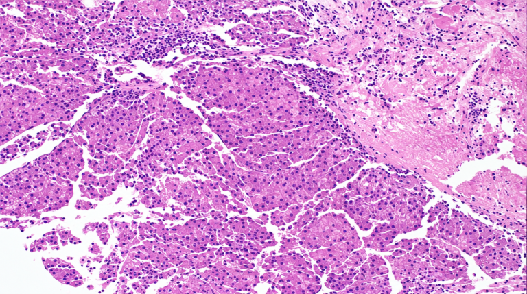
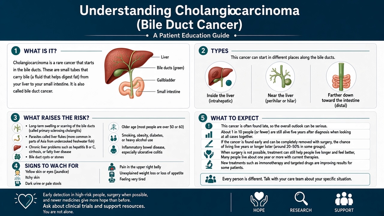
A Cholangiocarcinoma Pathology Report is the written medical interpretation of tissue taken from the bile ducts, liver hilum, gallbladder region, pancreas area, or nearby organs when bile duct cancer is suspected. Pathologists examine thin tissue sections under the microscope and decide whether the cells are benign, pre-cancerous, inflammatory, or malignant. When the diagnosis is cholangiocarcinoma, the report usually describes an adenocarcinoma, meaning a gland-forming cancer. The wording may also mention whether the tumor appears intrahepatic, perihilar, or distal, depending on where the cancer seems to arise.
A useful analogy is a building inspection after structural damage. Imaging may show where the building looks abnormal from the outside, but pathology examines the actual bricks and wiring. In the same way, a cholangiocarcinoma pathology report looks at the real tissue architecture and cell details. This is why pathology is central to confirming bile duct cancer rather than relying only on scans.
The report may include tumor grade, surgical margin status, lymph node involvement, and special testing. For patients still learning the basics, a general explanation of what a pathology report is can make this document easier to read. Tumor grade describes how abnormal the cancer cells look compared with normal bile duct lining. Surgical margin status describes whether cancer reaches the cut edge of the removed tissue. Lymph node involvement describes whether cancer is seen in sampled lymph nodes.
Why The Report Shows Cholangiocarcinoma
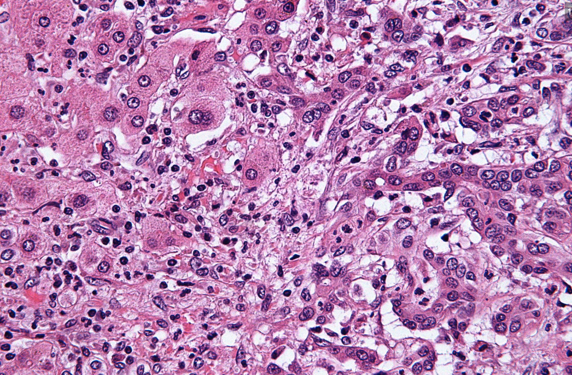
A diagnosis appears in the report because the pathologist saw malignant gland-forming cells with features consistent with bile duct cancer. These cells may form irregular glands, grow through tissue planes, produce dense scar-like stroma, or invade nerves and vessels. Perineural invasion means cancer cells are tracking around or along nerves, which is a common finding in cholangiocarcinoma. Vascular invasion means cancer cells are present inside blood vessels or lymphatic channels, which can affect staging and recurrence risk.
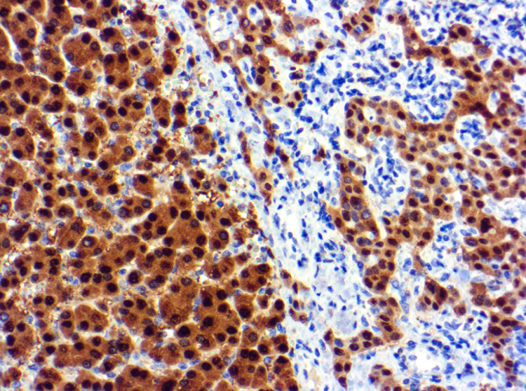
Sometimes the tissue sample is small, especially when the diagnosis comes from a needle biopsy, brush cytology, or endoscopic sampling. In small samples, the pathologist may use immunohistochemistry stains to support the diagnosis and help exclude cancers from other organs. Immunohistochemistry stains use antibodies to highlight proteins inside tumor cells, creating a pattern that can fit bile duct cancer or suggest another origin. These stains do not replace the microscope diagnosis, but they add important evidence when the appearance overlaps with pancreatic, gallbladder, stomach, colon, or metastatic cancers.
The report may also mention tumor grade, because some tumors form glands that still resemble bile duct lining, while others look much more disorganized. Surgical margin status may not appear if the sample was only a biopsy, because margins can usually be assessed only after a larger surgical specimen is removed. Lymph node involvement may also be absent from a biopsy report if no lymph nodes were submitted. For help understanding how different sections fit together, patients may benefit from reading a pathology report with confidence before an oncology visit.
How Serious Is A Cholangiocarcinoma Pathology Report?
A Cholangiocarcinoma Pathology Report is serious because cholangiocarcinoma is a malignant diagnosis, but the report does not automatically answer every question about prognosis. The seriousness depends on the tumor location, size, extent of spread, surgical margin status, lymph node involvement, tumor grade, imaging findings, liver function, and the patient’s overall health. Some cancers are found at a stage where surgery may be possible. Others are diagnosed when the tumor involves major bile ducts, blood vessels, liver tissue, or distant sites.
The cholangiocarcinoma pathology report should be read together with radiology and staging information. A small biopsy may confirm bile duct cancer but may not show the full depth of invasion, exact size, or all involved structures. Perineural invasion and vascular invasion can add concern because they show that cancer is using routes that may allow local spread or recurrence. Still, these findings need context, and they do not mean that treatment is pointless.
For a resection specimen, surgical margin status is one of the most closely reviewed lines. A negative margin means no cancer is seen at the cut edge examined by the pathologist, while a positive margin means cancer is present at that edge. Lymph node involvement is also important because positive nodes usually affect stage and may influence chemotherapy or radiation recommendations. Tumor grade helps describe aggressiveness, but grade alone is not the same as stage.
Patients often search the internet for survival numbers after seeing a cholangiocarcinoma pathology report, but broad statistics can be misleading for an individual person. The report gives essential facts, while the treating team combines those facts with scans, blood tests such as bilirubin and CA 19-9, procedure findings, and performance status. This is also a setting where expert review can matter, especially when the sample is small or the tumor origin is uncertain. A second review may confirm the diagnosis, refine the subtype, or recommend additional immunohistochemistry stains or molecular testing.
What Happens Next: Treatment And Monitoring
After a cholangiocarcinoma pathology report confirms bile duct cancer, the next step is usually a multidisciplinary plan. The team may include gastroenterology, hepatobiliary surgery, medical oncology, radiation oncology, interventional radiology, and pathology. Imaging studies help determine whether surgery is possible and whether the tumor is intrahepatic, perihilar, or distal. Blood work helps assess liver function and whether bile duct blockage needs drainage.
If surgery is performed, the final pathology report often contains more detail than the original biopsy. It may list tumor size, depth of invasion, tumor grade, surgical margin status, lymph node involvement, perineural invasion, and vascular invasion. Molecular testing may also be ordered, especially in advanced disease, because some cholangiocarcinomas have targetable alterations such as FGFR2 fusions or IDH1 mutations. Immunohistochemistry stains may also be used for mismatch repair proteins in selected cases.
Monitoring depends on the stage and treatment plan. Follow-up may include physical examinations, liver blood tests, tumor markers, and periodic imaging. For unresectable or metastatic bile duct cancer, treatment may include chemotherapy, immunotherapy, targeted therapy, radiation, stenting, or supportive procedures to improve bile flow. The pathology report remains the foundation document that connects the diagnosis to these treatment decisions.
Questions To Ask Your Doctor Or Pathologist
- Where does the tumor appear to arise: Intrahepatic, perihilar, distal bile duct, gallbladder region, pancreas, or another site?
- Does the report clearly confirm bile duct cancer, or are additional immunohistochemistry stains needed?
- What is the tumor grade, and how much should it influence treatment decisions?
- Was surgical margin status assessed, and are any margins positive or close?
- Was lymph node involvement found, and how many lymph nodes were examined?
- Does the report mention perineural invasion or vascular invasion?
- Should molecular testing be ordered for possible targeted therapy options?
These questions help turn a difficult report into a structured conversation. A cholangiocarcinoma pathology report contains many technical details, but each detail has a purpose. Patients may also ask whether the case was reviewed by a gastrointestinal pathologist or hepatobiliary pathology specialist.
A second opinion is especially reasonable when the diagnosis was made on a tiny biopsy, the tumor origin is uncertain, or treatment choices depend heavily on margin, node, or molecular findings. Honest Pathology explains when a second pathology opinion may be helpful and how expert review can clarify difficult reports. Another practical resource explains how to get a second opinion on a pathology diagnosis without starting from zero.
Frequently Asked Questions
Does cholangiocarcinoma on a pathology report always mean cancer?
Yes. Cholangiocarcinoma is a malignant tumor of the bile ducts, so a cholangiocarcinoma pathology report generally means bile duct cancer has been diagnosed in the sampled tissue. The remaining questions involve where the tumor started, how far it has spread, and which treatments are possible. A small biopsy may confirm cancer but not fully define stage.
Can a cholangiocarcinoma diagnosis be wrong?
Most diagnoses are reliable, but difficult cases can occur, especially with small samples or tumors near the pancreas, gallbladder, liver, stomach, or colon. Some cancers can look similar under the microscope. Immunohistochemistry stains, imaging correlation, and expert gastrointestinal pathology review can help confirm the origin. A second opinion may be appropriate if treatment depends on a very specific diagnosis.
What does positive margin mean in bile duct cancer?
A positive margin means cancer cells are present at the cut edge of the tissue removed during surgery. In a cholangiocarcinoma pathology report, surgical margin status helps the team judge whether visible disease may have been fully removed. A positive margin may affect recommendations for additional therapy. The meaning depends on which margin is involved and what treatments are available.
What does lymph node involvement mean in cholangiocarcinoma?
Lymph node involvement means cancer cells were found in one or more lymph nodes examined by the pathologist. This finding usually affects stage and may influence chemotherapy or radiation recommendations. The number of positive nodes and the total number examined both matter. Lymph node involvement is interpreted together with tumor size, location, margins, and imaging findings.
Should molecular testing be done for cholangiocarcinoma?
Molecular testing is often considered, particularly when bile duct cancer is advanced, recurrent, or not removable by surgery. A cholangiocarcinoma pathology report may mention whether enough tissue is available for testing. Results can sometimes identify targeted therapy options or immunotherapy-related markers. The oncology team can explain which tests are appropriate for the specific clinical situation.
A cholangiocarcinoma pathology report can feel like a door closing, but it is also the document that helps open the next steps in care. The most helpful path forward is careful review, clear questions, and correlation between pathology, imaging, and clinical findings. Honest Pathology consultations can provide an expert explanation or second review when patients and families need the report translated into plain language.
References:
National Cancer Institute — Cholangiocarcinoma Definition
National Cancer Institute — Pathology Reports Fact Sheet
National Cancer Institute — Second Opinion Definition