Finding out you’ve been diagnosed with skin cancer can feel overwhelming , and reading the pathology report that comes with it often adds to the confusion. The report is full of technical words, microscopic descriptions, and staging details that may seem impossible to understand without a medical degree.
But here’s the truth: you deserve to understand your diagnosis clearly. Every word in that report tells a story about your skin cells , where the cancer started, how deep it goes, and what type it is. Once you understand what your pathology report means, you can have more productive conversations with your dermatologist, surgeon, or oncologist and feel confident in your next steps.
That’s exactly why Honest Pathology™ exists , to help patients interpret their reports in plain language and feel empowered by knowledge, not overwhelmed by jargon.
What Is a Pathology Report for Skin Cancer
A pathology report is a detailed document written by a pathologist , a medical doctor who specializes in diagnosing disease by examining tissue under a microscope.
When your dermatologist removes a mole, lesion, or suspicious spot on your skin, that sample is sent to a pathology laboratory. The pathologist processes the tissue, stains it, and studies it under the microscope. They look for abnormal cells, patterns, and other signs that tell whether it’s benign (non-cancerous), pre-cancerous, or malignant (cancerous).
Their findings are summarized in your skin pathology report, which becomes part of your medical record. This report plays a key role in guiding your treatment. It helps determine whether additional surgery is needed to remove more tissue, whether specialized options such as Mohs surgery, immunotherapy, or radiation would be appropriate, and whether your doctor should examine nearby lymph nodes.
The Major Types of Skin Cancer You Might See in a Pathology Report
Not all skin cancers are the same. Your pathology report will specify what type of skin cancer was found. The three most common are:
1. Basal Cell Carcinoma (BCC)
The most common type of skin cancer, BCC develops from the basal cells at the bottom of the epidermis. It usually grows slowly and rarely spreads to other parts of the body. However, if left untreated, it can cause significant local tissue damage.
Your report might include terms like “nodular,” “superficial,” or “infiltrative,” describing the growth pattern of the tumor.
2. Squamous Cell Carcinoma (SCC)
SCC arises from the squamous cells that make up most of the skin’s outer layer. It can spread (metastasize) if not completely removed, especially when deeply invasive or poorly differentiated.
Pathologists often include words like “well-differentiated” or “poorly-differentiated,” which describe how aggressivethe tumor cells look.
3. Melanoma
Melanoma originates in melanocytes, the cells that produce pigment (melanin). Though less common, melanoma can be much more dangerous because it can spread quickly to lymph nodes or distant organs.
Melanoma pathology reports are often the most detailed, with specific measurements and terms that affect staging and treatment decisions.

Key Sections of a Skin Cancer Pathology Report
Let’s break down the main sections of your report so you can understand what they mean in plain language.
1. Clinical Information
This section includes background information from your dermatologist , such as the body site, size, and appearance of the lesion.
Example: “Shave biopsy of a pigmented lesion, left forearm.”
2. Gross Description
This is what the sample looked like to the naked eye , its color, size, and shape.
Example: “Tan-brown skin ellipse measuring 1.0 x 0.6 cm.”
This part is mostly technical and doesn’t influence your diagnosis directly.
3. Microscopic Description
Here’s where the pathologist describes what they saw under the microscope. Expect scientific terms describing the cells’ shape, arrangement, and depth.
Example: “Infiltrative nests of basaloid cells extending into the dermis.”
While this language is meant for doctors, it contains clues about how advanced or aggressive the cancer might be.
4. Diagnosis or Final Diagnosis
This section is often the most important for patients because it summarizes the findings in a clear, direct statement. For example, your report might read, “Basal cell carcinoma, nodular type, completely excised,” or “Invasive squamous cell carcinoma, moderately differentiated,” or “Superficial spreading melanoma, Breslow depth 1.2 mm.” This single line captures the diagnosis in a simple, straightforward way.
That short paragraph defines the type of skin cancer, growth pattern, and sometimes whether the entire lesion was fully removed.
Special Terms You’ll See in Skin Cancer Reports , Explained in Plain Language
Understanding a few key terms can make your report much easier to read:
| Term | Meaning |
| Margins | The edges of the removed tissue. If “clear” or “negative,” it means no cancer was seen at the edges , good news. If “positive,” cancer cells extend to the edge, and your doctor may need to remove more tissue. |
| Invasive | The cancer has grown beyond the surface layer of the skin into deeper layers. |
| In situ | Cancer cells are present but confined to the top layer (epidermis) , early and highly curable. |
| Breslow Depth | Used for melanoma , the thickness of the tumor in millimeters, which helps determine stage and prognosis. |
| Ulceration | A breakdown of the skin over the tumor. Presence of ulceration can make melanoma more aggressive. |
| Mitotic Rate | How many cells are dividing. A higher number suggests a faster-growing tumor. |
| Lymphovascular Invasion | Whether cancer cells are seen inside blood or lymph vessels , a sign that the cancer could spread. |
| Perineural Invasion | Cancer cells around nerves. Important in SCC because it increases the risk of recurrence. |
Each of these terms helps your doctor build a picture of how serious the cancer is and what kind of treatment you might need next.
How Pathologists Determine “Stage” and “Grade”
For melanoma and some squamous cell carcinomas, your report may mention staging.
- Stage tells how far the cancer has spread (thickness, ulceration, and sometimes lymph node involvement).
- Grade describes how “abnormal” the cells look compared to normal skin cells. The more abnormal, the higher the grade , and the more likely the cancer may behave aggressively.
If your report doesn’t include a stage, don’t panic , sometimes pathologists can’t assign a full stage until after surgical excision and lymph node evaluation.
Why Margins Matter So Much
One of the first things patients want to know is, “Did they get it all?”
Your pathology report will have a section about surgical margins, meaning whether cancer cells reach the outer edge of the removed tissue.
- Negative margins = all cancer removed.
- Positive margins = some cancer cells remain at the edge, so your doctor may recommend another excision.
Sometimes, you’ll see “close margins,” meaning cancer came within a fraction of a millimeter of the edge. Your care team will discuss whether further treatment is needed.
What If My Pathology Report Says “Atypical” or “Precancerous”?
Not every abnormal skin biopsy is cancer. Sometimes, the pathologist reports findings such as:
- Actinic keratosis (pre-cancerous change caused by sun damage)
- Atypical nevus or dysplastic nevus (a mole with irregular cells but not melanoma)
These lesions are often removed to prevent progression to cancer. Honest Pathology frequently helps patients understand these “borderline” findings , the kind that aren’t clearly cancer but still cause worry.
Understanding exactly what “atypical” means in your specific context can give peace of mind and guide smart follow-up with your dermatologist.
How to Read Your Report Without Fear
When you first see your pathology report, you might feel anxious. That’s completely normal. Here are a few ways to approach it calmly:
- Start with the “Final Diagnosis” section. That’s the summary of what the sample showed.
- Look for words like “in situ,” “invasive,” and “margins.” These reveal the stage and completeness of removal.
- Note any mention of subtype or depth. Especially for melanoma, the Breslow depth is key.
- Don’t fixate on scary words. Terms like “mitotic rate” or “ulceration” are important for doctors but don’t define your prognosis alone.
- Ask your dermatologist to explain anything unclear. Pathologists and dermatologists work together , you deserve clear answers.
And if you’re still uncertain, that’s where Honest Pathology™ can help.
How Honest Pathology™ Helps You Understand Your Skin Cancer Diagnosis
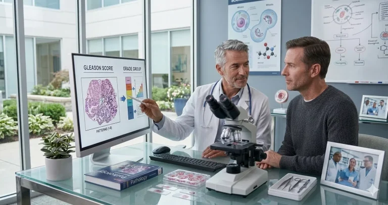
At Honest Pathology, our mission is simple: to make pathology understandable. We believe every patient deserves to know exactly what their report means in plain, caring, and accurate language. When you connect with an Honest Pathologist, you receive a one-on-one educational consultation over a secure, HIPAA-compliant platform. Your pathologist walks you through your report line by line using everyday terms, often with visual aids and microscope images that show what was seen during the examination. You’ll gain clarity about what is confirmed, what remains uncertain, and which key questions you can bring back to your doctor.
The goal is peace of mind knowing you fully understand your diagnosis and what happens next. These consultations are educational only; we don’t provide second opinions or treatment recommendations. Instead, we empower you to understand your report so you can communicate with confidence and make informed decisions about your care.
Why Understanding Your Pathology Report Matters
Knowledge is power, and in medicine, it’s also protection. When patients truly understand their pathology reports, they’re able to make informed decisions about surgery or follow-up, spot errors or inconsistencies early, ask stronger questions during appointments, and reduce the fear that comes from not knowing what a diagnosis really means.
Many patients tell us that after meeting with Honest Pathology, they felt calmer, more confident, and prepared to face treatment decisions head-on. Understanding turns fear into clarity and clarity into strength.
When to Ask for a Second Pathology Review
Sometimes, doctors recommend a second pathology opinion, especially when a diagnosis is unusual or rare, when the report includes phrases like “borderline” or “cannot rule out,” or when the results could change treatment decisions, such as distinguishing melanoma from a dysplastic nevus.
While Honest Pathology doesn’t provide formal second opinions, our consultations can help you understand when seeking one might be helpful. We review your report carefully and explain whether the wording or findings indicate that another expert’s review could be beneficial.
Final Thoughts
Your skin cancer pathology report is not just paperwork , it’s the scientific story of your skin cells, written by a specialist who studies disease under the microscope. The challenge is that it’s written for other doctors, not for you.
At Honest Pathology™, we bridge that gap. We help you read your report, understand your diagnosis, and ask the right questions , all in plain language and in the comfort of your own home. You’ll come away not just with knowledge, but with the confidence that comes from truly understanding your own health.
If you’ve recently received a skin biopsy or cancer report and want to know exactly what it means, visit HonestPathology.com to schedule your educational consultation today. Because knowledge isn’t just power, it’s peace of mind.