
Worried after colonoscopy? tubular vs tubulovillous adenoma explains polyp risk, dysplasia, and what to ask next.
tubular vs tubulovillous adenoma usually describes two common precancerous colon polyp patterns, not a diagnosis of invasive cancer. These words come from how the polyp looks under the microscope after a colonoscopy biopsy or polyp removal. The distinction helps doctors estimate future colorectal cancer risk and decide when the next colonoscopy should happen.
Seeing the word “adenoma” on a colon polyp report can feel frightening, especially when the report also mentions dysplasia, villous features, or margins. Many patients hear “precancerous” and immediately imagine the worst. In pathology, however, these terms are meant to sort risk carefully, not to label every polyp as dangerous.
A colon polyp report is like a maintenance report after finding a weak spot in a bridge. The report does not mean the bridge has collapsed; it explains what type of weak spot was found, how advanced it looked, and whether the area appears to have been fully removed. Clear interpretation often turns fear into a focused plan.
Tubular Vs Tubulovillous Adenoma — What It Actually Means
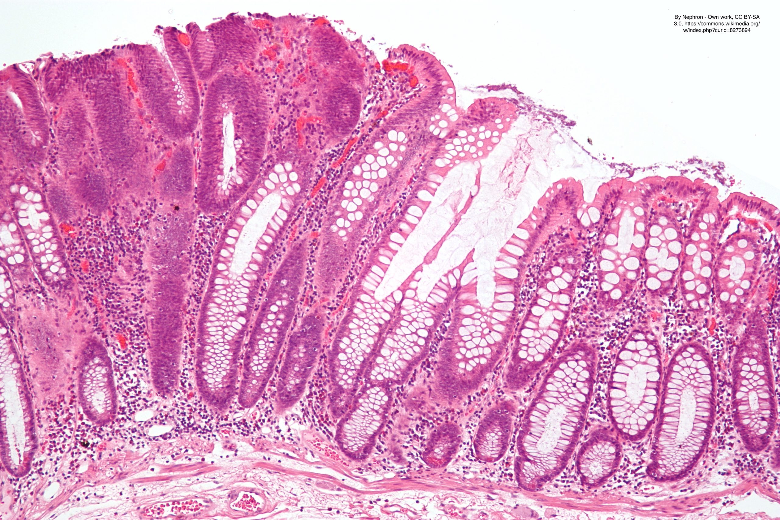
The phrase tubular vs tubulovillous adenoma refers to the microscopic architecture of an adenomatous colon polyp. A tubular adenoma is made mostly of small tube-like glands, which is the most common and generally lower-risk adenoma pattern. A tubulovillous adenoma contains a mixture of tubular glands and villous features, which are longer, finger-like projections. Pathologists use these patterns because the amount of villous growth can affect the recommended follow-up interval.
A helpful analogy is the shape of a carpet. A tubular adenoma is more like a short, even pile, while a tubulovillous adenoma has some shaggy, projecting areas. The shaggy areas are not cancer by themselves, but they can be associated with a higher chance of advanced precancerous change. That is why villous features are often highlighted in a colon polyp report.
The diagnosis is made by a pathologist who examines thin tissue sections from the colonoscopy biopsy or removed polyp. The report may also list size, number of polyps, adenoma dysplasia grade, and whether the polyp margins are clear. These details matter together; no single word tells the whole story. Patients who want a calmer starting point for report language may also find What Is a Pathology Report? helpful before reviewing the details with a clinician.
Why The Report Shows This Finding
Adenomas form when cells lining the colon begin growing in a more organized but abnormal way. The abnormal growth can develop slowly over years, which is why screening colonoscopy is so effective. Many adenomas are found before symptoms appear and before colorectal cancer develops. A colonoscopy biopsy gives the laboratory a sample that can be classified by architecture and dysplasia.
The adenoma dysplasia grade describes how abnormal the cells look, not whether the polyp has already become invasive cancer. Most adenomas have low-grade dysplasia, meaning the cells are abnormal but still limited to the lining pattern expected in an adenoma. High grade dysplasia means the cells look more advanced and crowded, but it still does not automatically mean invasive colorectal cancer. Pathologists look for true invasion into deeper tissue before using the word cancer.
Villous features may appear because part of the adenoma grows with elongated surface projections rather than simple tubes. Larger polyps are more likely to show villous features, high grade dysplasia, or more complex architecture. Polyp margins are evaluated when the specimen is removed in a way that allows assessment of whether adenomatous tissue reaches the edge. In many small fragmented polyps, polyp margins may not be assessable, and that limitation is a common technical issue rather than a sign that something was missed.
How Serious Is Tubular Vs Tubulovillous Adenoma?
The seriousness of tubular vs tubulovillous adenoma depends on the full set of findings, especially polyp size, number, dysplasia, completeness of removal, and prior colonoscopy history. A small tubular adenoma with low-grade dysplasia and complete removal is usually considered a lower-risk finding. A larger tubulovillous adenoma, especially one with high grade dysplasia, usually needs closer follow-up. The key message is that these findings describe risk, not certainty.
In real-world terms, tubular vs tubulovillous adenoma is a warning light, not a crash. The warning light is useful because it allows doctors to act early by removing the polyp and planning surveillance. Tubulovillous adenoma has more villous features than a purely tubular adenoma, and that is why it may carry a higher future risk if similar polyps continue to develop. Still, once the polyp has been completely removed, the immediate problem may already have been treated.
Adenoma dysplasia grade is one of the most important lines in the pathology report. Low-grade dysplasia is expected in most conventional adenomas, while high grade dysplasia suggests a more advanced precancerous change. Polyp margins can also influence the plan, particularly for large polyps removed in pieces or removed by advanced endoscopic techniques. When a colon polyp report contains multiple higher-risk features, the gastroenterologist may recommend an earlier repeat colonoscopy to confirm that the area is clear.
What Happens Next: Treatment And Monitoring
For most patients, the treatment has already happened during colonoscopy because the polyp was removed. The next step is usually surveillance, meaning a planned repeat colonoscopy at an interval based on the complete findings. Doctors consider the colon polyp report together with the endoscopy report, bowel preparation quality, family history, and prior polyps. A colonoscopy biopsy result is only one part of that decision.
Follow-up timing is not the same for every adenoma. Small tubular adenomas may lead to a longer interval before the next colonoscopy, while larger adenomas, multiple adenomas, tubulovillous adenoma, high grade dysplasia, or uncertain polyp margins may lead to closer surveillance. This does not mean cancer is expected. It means the care team wants to prevent colorectal cancer by checking again at the right time.
If the wording feels confusing, a structured review can help. The report should be compared with the procedure note because the pathologist may not know whether the endoscopist thought the polyp was fully removed. Patients trying to understand each line may benefit from Understanding Your Pathology Report: How to Read It with Confidence. When the diagnosis includes unusual wording, difficult margins, or high-risk features, a second review may also be reasonable.
Questions To Ask The Doctor Or Pathologist
- Was the polyp a tubular adenoma, a tubulovillous adenoma, or another type of polyp?
- What was the adenoma dysplasia grade, and was high grade dysplasia present?
- How large was the polyp, and how many adenomas were found during colonoscopy?
- Were villous features present, and approximately how much of the polyp showed them?
- Were the polyp margins clear, involved, or not possible to assess?
- Was the colonoscopy biopsy or polyp removal considered complete by the endoscopist?
- When should the next colonoscopy be scheduled based on the full report?
These questions help connect the microscope findings with the procedure findings. Pathologists often classify the tissue pattern, while gastroenterologists decide the surveillance interval using the full clinical picture. A patient does not need to memorize pathology terminology to have a productive discussion.
A second opinion is most useful when the diagnosis may change management, such as suspected cancer, high grade dysplasia, difficult polyp margins, or uncertainty about villous features. Helpful information about timing and reasons for review is available in When Should You Get a Second Pathology Opinion?. For patients already gathering slides and reports, How to Get a Second Opinion on Your Pathology Diagnosis explains the practical steps.
Frequently Asked Questions
Is a tubulovillous adenoma cancer?
A tubulovillous adenoma is usually not cancer. It is a precancerous adenoma pattern that has some villous features, which can carry a higher risk than a small tubular adenoma. The report should be checked for high grade dysplasia or invasive carcinoma because those terms change the level of concern. If the polyp was completely removed, the main next step is usually surveillance rather than cancer treatment.
Is a tubular adenoma less serious than a tubulovillous adenoma?
In general, a tubular adenoma is often lower risk than a tubulovillous adenoma, especially when it is small and has low-grade dysplasia. The phrase tubular vs tubulovillous adenoma is helpful because it separates common lower-risk architecture from architecture with more villous growth. Size, number of polyps, adenoma dysplasia grade, and removal status still matter. A small tubulovillous adenoma may be less concerning than a very large tubular adenoma with other high-risk features.
What does low grade dysplasia mean in a colon polyp?
Low-grade dysplasia means the polyp cells look abnormal under the microscope but do not show the most advanced precancerous changes. Most conventional adenomas have low-grade dysplasia by definition. This wording does not mean invasive colorectal cancer is present. The doctor will still use the colon polyp report to choose the safest follow-up interval.
What if my polyp margins are positive?
Positive polyp margins mean adenoma tissue extends to the cut edge of the submitted specimen. This can mean residual polyp may remain, but interpretation depends on how the polyp was removed and whether the specimen was fragmented. The gastroenterologist may recommend repeat colonoscopy or close inspection of the removal site. Polyp margins are most meaningful when the specimen is intact and oriented well enough for assessment.
Can tubular vs tubulovillous adenoma change after a second opinion?
Yes, tubular vs tubulovillous adenoma can sometimes be refined after expert review, especially when villous features are borderline or the sample is fragmented. Pathologists may also recheck the adenoma dysplasia grade and look carefully for high grade dysplasia or invasion. Many reviews confirm the original diagnosis, which can still provide reassurance. A change matters most when it affects surveillance timing or the need for additional treatment.
A report mentioning tubular vs tubulovillous adenoma can sound intimidating, but it often represents a preventable-risk finding that has already been removed. The safest next step is to connect the pathology details with the colonoscopy findings and the recommended surveillance plan. Honest Pathology consultations can help clarify confusing wording, confirm key risk features, and support a more confident discussion with the treating doctor.
References:
National Cancer Institute — Pathology Reports
National Cancer Institute — Definition of Adenoma
MedlinePlus — Colorectal Cancer
