

A STIC fallopian tube result can feel frightening. Learn what this finding means, why it matters, and what to ask the doctor next.
STIC fallopian tube means that a pathologist found a very early abnormal change in the lining cells of the fallopian tube, most often near the end closest to the ovary. STIC stands for serous tubal intraepithelial carcinoma, a diagnosis that is usually made under the microscope after surgery, not by symptoms alone. It is closely linked to the pathway that can lead to high grade serous carcinoma, the most common and aggressive type of ovarian, fallopian tube, and primary peritoneal cancer.
Seeing the word carcinoma in a report can feel like the floor has dropped away. Many patients discover this term after a risk-reducing surgery, hysterectomy, or removal of ovaries and tubes, when no one expected a cancer-related finding. The clearest first step is to understand exactly what was found, whether anything invasive was present, and what follow-up is recommended.
A pathology report is not just a label; it is a map of what the tissue showed. Helpful background on the structure of these reports is available in What Is a Pathology Report?, especially for patients seeing unfamiliar terms for the first time.
STIC Fallopian Tube — What It Actually Means
STIC is short for serous tubal intraepithelial carcinoma. In plain language, it means that the abnormal cells are sitting within the surface lining of the fallopian tube rather than clearly growing into deeper tissue. The word intraepithelial is the key part: It tells the clinical team that the abnormality is confined to the lining layer in the examined section. That distinction matters because serous tubal intraepithelial carcinoma is not the same as a large invasive tumor.
A useful analogy is damaged paint on the surface of a wall versus rot that has spread into the wooden frame. STIC fallopian tube describes a surface-level cellular abnormality, while invasive carcinoma means abnormal cells have broken beyond their original layer and entered deeper tissue. Pathologists carefully examine the fallopian tube, especially the fimbrial end, because many high grade serous carcinoma cases are now believed to begin there. This is why fallopian tube pathology has become so central in gynecologic cancer prevention and diagnosis.
Under the microscope, the cells in serous tubal intraepithelial carcinoma usually look crowded, irregular, and more atypical than normal fallopian tube lining cells. The pathologist may use immunohistochemistry, especially p53 staining, to support the diagnosis. Many reports also mention Ki-67, a marker of cell growth; patients who see that term may find What Does Ki-67 Mean and How High Is Concerning? helpful for general context. STIC fallopian tube is therefore a microscopic diagnosis that carries important risk information, even when no mass was seen during surgery.
Why Your Report Shows This Finding
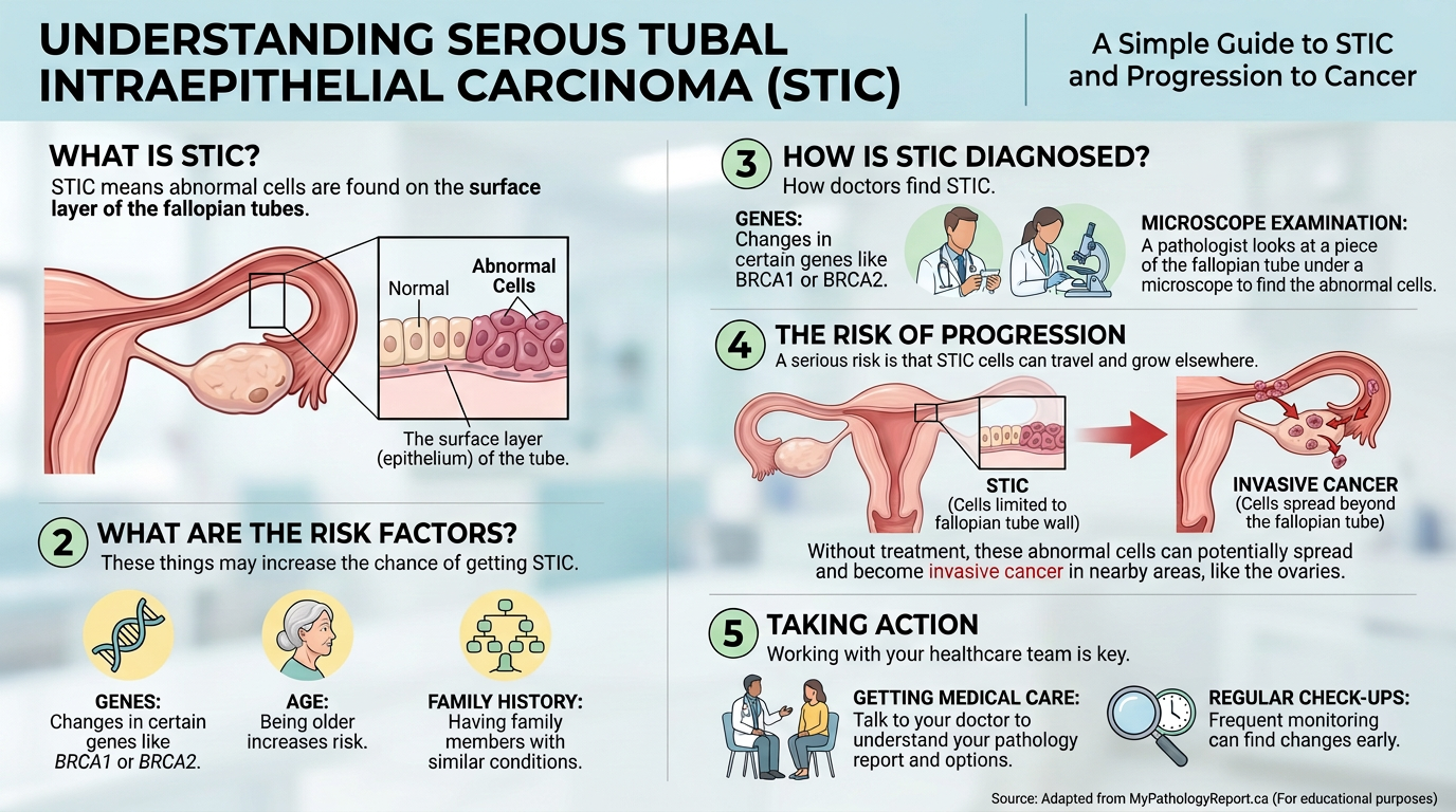
A report may show STIC because the fallopian tubes were removed and processed carefully, often using a method that examines the fimbrial end in detail. This can happen during risk-reducing surgery for a BRCA gene mutation, surgery for another gynecologic condition, or staging surgery for a known cancer. The finding may be unexpected because STIC often does not form a visible lump. In many cases, the surgeon sees no obvious abnormality, and the diagnosis comes entirely from fallopian tube pathology.
Pathologists look for a combination of abnormal cell shape, abnormal growth pattern, and supportive special stains. Strong or completely absent p53 staining can suggest an underlying TP53 abnormality, which is common in the pathway toward high grade serous carcinoma. A high Ki-67 growth pattern may also support serous tubal intraepithelial carcinoma when the cell appearance fits. These details help separate STIC from benign reactive changes, which can sometimes look worrisome but do not carry the same meaning.
The clinical context is also important. A BRCA gene mutation, especially BRCA1 or BRCA2, increases lifetime risk for ovarian and related cancers, and STIC may be found during preventive removal of tubes and ovaries. Some pathology reports also mention pelvic washings, which are fluid samples collected from the abdomen or pelvis to look for abnormal cells. Pelvic washings can help determine whether abnormal serous cells are floating elsewhere in the pelvic cavity. When STIC fallopian tube is reported, the care team usually reviews the tubes, ovaries, omentum if sampled, pelvic washings, and any other tissue submitted at surgery.
How Serious Is STIC Fallopian Tube?
STIC fallopian tube is serious because it is considered a precursor or very early form in the high grade serous carcinoma pathway. That does not automatically mean that an invasive cancer is present. The seriousness depends on whether the abnormality is isolated, whether invasive carcinoma is found anywhere, whether pelvic washings are positive, and whether the patient has a BRCA gene mutation or other hereditary risk. In an isolated STIC, the report may show no tumor mass and no invasion, which is very different from advanced cancer.
The most reassuring scenario is an isolated serous tubal intraepithelial carcinoma with completely examined ovaries and tubes, no invasive carcinoma, and negative pelvic washings. Even then, many gynecologic oncologists recommend careful follow-up because STIC may signal increased risk for later primary peritoneal carcinoma. A more concerning scenario is STIC fallopian tube found together with invasive high grade serous carcinoma in the ovary, tube, peritoneum, or omentum. In that situation, the STIC may represent the starting point or a related finding, while the invasive cancer drives staging and treatment.
Patients often ask whether STIC is cancer or pre-cancer, and the answer is nuanced. The term serous tubal intraepithelial carcinoma includes the word carcinoma because the cells show high-grade malignant features, but they are confined to the epithelial lining in the area examined. In practical care, STIC fallopian tube is treated as a high-risk pathologic finding rather than ignored as a minor abnormality. A second pathology review can be especially valuable if the diagnosis was unexpected, if p53 staining was borderline, or if treatment decisions depend on whether invasion is truly absent.
What Happens Next: Treatment and Monitoring
Next steps usually involve a gynecologic oncologist, even when no invasive tumor is identified. The doctor may review whether the original surgery removed both fallopian tubes and ovaries, whether staging samples were taken, and whether pelvic washings were performed. If surgery was limited, additional staging may be discussed depending on age, risk factors, operative findings, and the exact fallopian tube pathology. The goal is not to create panic; the goal is to make sure no associated invasive disease was missed.
Monitoring may include physical exams, symptom review, and sometimes CA-125 blood testing or imaging, although follow-up plans vary. For patients with a BRCA gene mutation, genetic counseling and family risk assessment may also be part of care. Relatives may need information about hereditary cancer testing, but that discussion should be guided by genetics professionals. High grade serous carcinoma risk management is most effective when pathology, surgery, genetics, and oncology are considered together.
Some patients are told that observation is appropriate, while others are offered additional surgery or closer surveillance. The difference often comes down to whether STIC was isolated, whether ovaries remain, whether pelvic washings were positive, and whether there is a hereditary cancer syndrome. Patients trying to understand the wording of the report may benefit from Understanding Your Pathology Report: How to Read It with Confidence. STIC fallopian tube is a diagnosis where the small details in the pathology report can meaningfully shape the next conversation.
Questions to Ask Your Doctor or Pathologist
- Was the STIC isolated, or was any invasive high grade serous carcinoma found anywhere?
- Which part of the tube contained the abnormality, especially the fimbrial end?
- Were both fallopian tubes and ovaries completely examined by the pathology team?
- What did the p53 staining and Ki-67 results show, and did they strongly support the diagnosis?
- Were pelvic washings performed, and were any abnormal cells found?
- Should genetic counseling or testing for a BRCA gene mutation be considered?
- Would a second pathology opinion change staging, follow-up, or treatment recommendations?
These questions are specific because STIC is a microscopic diagnosis with real clinical consequences. A patient does not need to memorize pathology terminology, but clear answers can reduce uncertainty and help the next appointment feel more focused.
A second review is often reasonable when a rare or high-impact diagnosis appears in a report. More information is available in When Should You Get a Second Pathology Opinion?. For STIC fallopian tube, that review typically checks the original slides, the p53 staining pattern, the presence or absence of invasion, and whether any other submitted tissue shows related disease.
Frequently Asked Questions
Is STIC in the fallopian tube cancer?
STIC is a high-grade abnormality in the lining of the fallopian tube and is called serous tubal intraepithelial carcinoma. It is not the same as a bulky invasive cancer when the abnormal cells remain confined to the lining. STIC fallopian tube still matters because it is strongly linked to the pathway that can lead to high grade serous carcinoma. The most important question is whether the report found any invasive carcinoma elsewhere.
Can STIC spread if it is only in the tube?
An isolated STIC does not show invasion in the tissue section where it was diagnosed. However, abnormal serous cells may sometimes be associated with positive pelvic washings or later peritoneal disease, which is why follow-up is usually recommended. Doctors consider the entire surgical and pathology picture, not the STIC line alone. Negative pelvic washings and no invasive cancer are generally more reassuring findings.
Does STIC mean ovarian cancer?
STIC does not automatically mean ovarian cancer is present. It means that serous tubal intraepithelial carcinoma was found in the fallopian tube lining. Because many high grade serous carcinoma cases involving the ovary or peritoneum may begin near the fimbrial end of the tube, the finding is taken seriously. The ovaries, tubes, and any other submitted tissues must be reviewed before conclusions are made.
Should pathology slides be reviewed for STIC?
A second pathology opinion can be helpful because STIC fallopian tube is uncommon and can affect follow-up decisions. Review usually focuses on whether the abnormal cells truly meet criteria for STIC, whether p53 staining supports the diagnosis, and whether there is any invasion. It may also confirm whether other tissue samples show high grade serous carcinoma. Guidance on the process is available in How to Get a Second Opinion on Your Pathology Diagnosis.
What follow-up is needed after STIC?
Follow-up depends on whether STIC was isolated, whether surgery was complete, whether pelvic washings were negative, and whether a BRCA gene mutation is present. Many patients are followed by a gynecologic oncologist with periodic exams and individualized testing. Some may need additional staging or genetic counseling. STIC fallopian tube should prompt a careful plan, but it does not always mean chemotherapy is needed.
A STIC diagnosis can feel overwhelming, especially when it appears after surgery that was expected to be preventive or routine. Clear pathology review can separate an isolated microscopic finding from invasive disease and give the care team a stronger basis for decisions. Honest Pathology consultations can help patients and families understand what the report says, what it does not say, and which questions deserve attention next.
References:
National Cancer Institute — Pathology Reports
National Cancer Institute — Fallopian Tube Cancer
National Cancer Institute — BRCA1 Gene
