
Seeing STAS can feel frightening. STAS lung cancer affects risk discussions, surgery choices, and follow-up. Learn what to ask next.
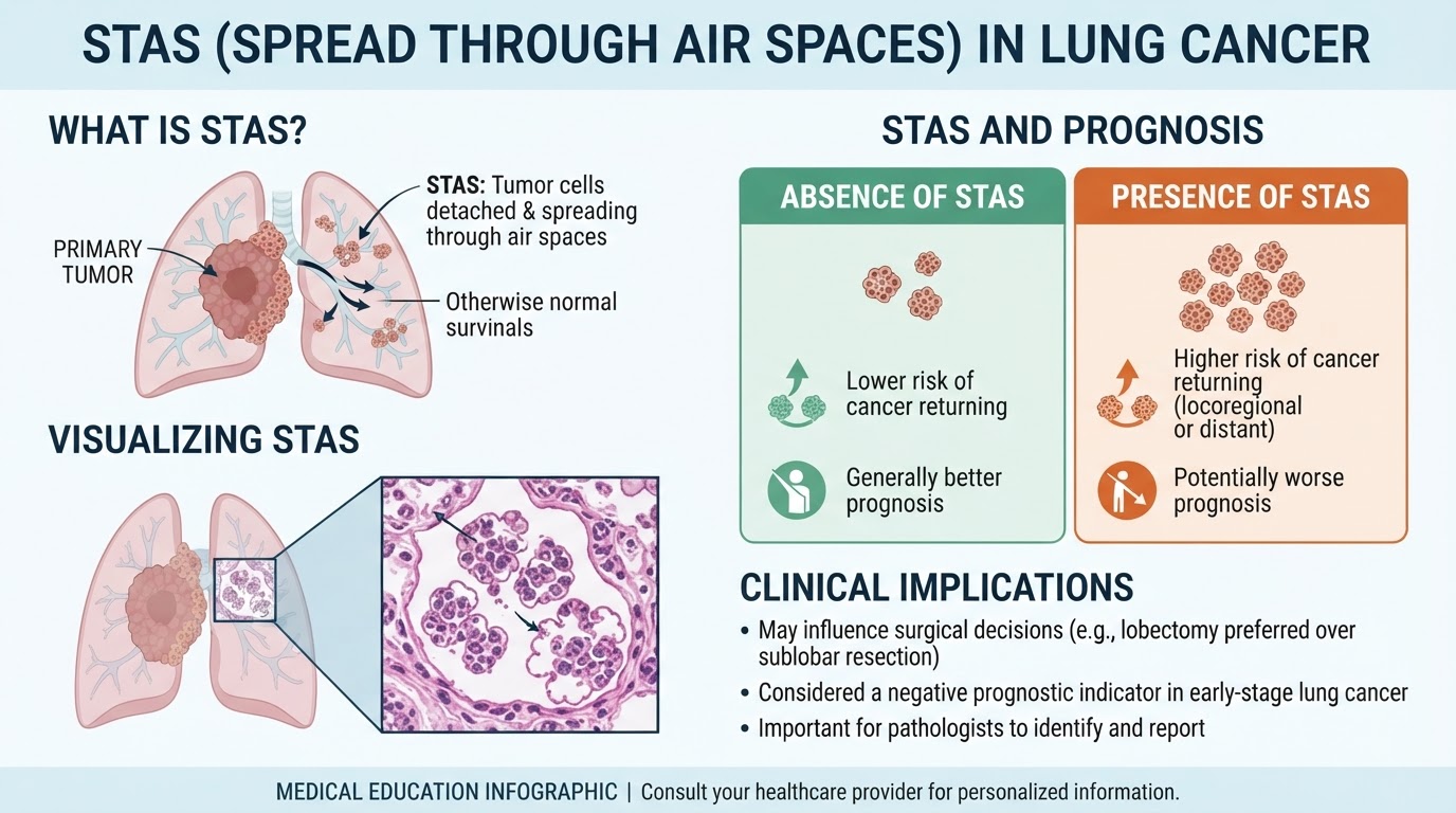
STAS lung cancer means that pathologists saw cancer cells spreading into nearby air spaces in the lung, separate from the main tumor. This finding is most often discussed in non-small cell lung cancer, especially lung adenocarcinoma, and it can affect how the cancer team thinks about surgery, recurrence risk, and follow-up. STAS does not automatically mean the cancer has spread throughout the body, and it is not the same as metastatic disease.
Seeing an unfamiliar term on a pathology report can make an already stressful diagnosis feel even heavier. Many patients search the term late at night because it sounds technical, ominous, and unexplained. Clear language matters because STAS is a microscopic finding, not a sentence or a prediction by itself.
A pathology report is best understood as a map. STAS is one landmark on that map, along with tumor size, lymph nodes, surgical margin status, tumor type, grade, and stage. The goal is to understand what this landmark may mean in the full clinical picture.
STAS Lung Cancer — What It Actually Means
STAS lung cancer refers to “spread through air spaces,” a pattern seen under the microscope when clusters, strands, or single tumor cells are present in air-filled spaces of the lung beyond the edge of the main tumor. In plain language, the main tumor is like an island, and STAS describes small groups of cancer cells floating in the nearby air channels around that island. These cells are not seen by touch or by a routine scan; they are identified by examining thin tissue sections on glass slides.
Pathologists most often report STAS in lung adenocarcinoma, although it can also be described in other types of lung cancer. The finding is part of a detailed pathology report after a biopsy or, more commonly, after a lung resection. A small biopsy may not show enough surrounding lung tissue to evaluate STAS reliably, so the term is usually most meaningful after surgery. For patients still learning the basics, what a pathology report contains can provide helpful context before focusing on one specific line.
The key idea is that STAS is a local microscopic growth pattern. It does not mean that cancer has entered the bloodstream, and it does not prove that lymph nodes contain cancer. It does, however, suggest that tumor cells have a tendency to extend beyond the visible border of the mass. That is why STAS lung cancer can become part of discussions about the surgical margin, type of operation performed, and the risk of cancer recurrence.
Why Your Report Shows This Finding
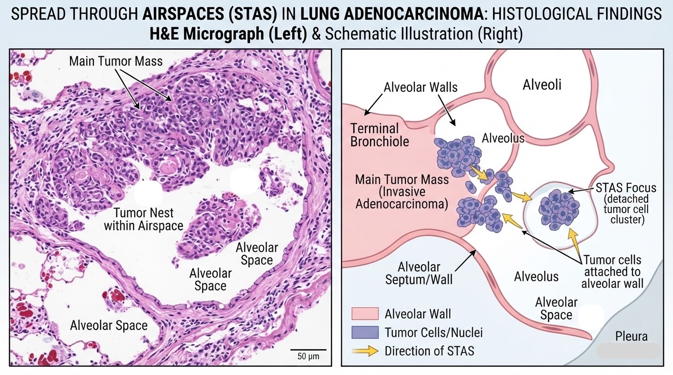
A pathology report shows STAS when the pathologist sees tumor cells in alveolar spaces away from the main tumor edge. Alveoli are tiny air sacs where oxygen exchange happens, and cancer cells can sometimes grow or detach into these delicate spaces. The medical phrase spread through air spaces describes this pattern. It is a microscopic observation made after the tissue is processed, stained, and reviewed slide by slide.
Several tumor features may travel together with STAS, especially in lung adenocarcinoma. Some tumors have more fragile or discohesive tumor cells, meaning the cells separate from one another more easily. Certain growth patterns, higher grade features, lymphovascular invasion, or a larger tumor size may also raise concern, although every case must be interpreted individually. The pathologist does not diagnose STAS from a blood test or imaging study; it is a tissue-based finding.
There are also technical issues that pathologists consider carefully. During tissue handling, some tumor cells can become displaced, creating an artifact that can mimic true spread through air spaces. Experienced lung pathologists look at the overall pattern, location, cell clusters, and relationship to the main tumor before reporting STAS. This is one reason a second review may be useful when a pathology report contains a finding that could influence treatment. A patient trying to understand several report elements together may benefit from learning how to read a pathology report with confidence.
How Serious Is STAS Lung Cancer?
STAS lung cancer is serious enough to discuss with the treating team, but it should not be interpreted in isolation. Research has linked STAS with a higher risk of cancer recurrence in some patients, particularly when smaller lung cancers are treated with limited surgery such as wedge resection. The reason is intuitive: if tumor cells are present beyond the main mass, removing only a narrow rim of nearby lung may leave less room for safety. Still, the actual risk depends on tumor stage, lymph node status, surgical margin distance, tumor type, and the operation performed.
For some patients, STAS lung cancer may have little practical effect because the tumor was removed by lobectomy with clear margins and lymph nodes were negative. For others, especially patients with early lung adenocarcinoma treated by sublobar resection, STAS can be more relevant to decisions about surveillance or additional therapy. A clear surgical margin remains reassuring, but the margin must be interpreted alongside the microscopic growth pattern. If the surgical margin is close or if the specimen is small, the care team may review whether the surgery achieved adequate local control.
The most helpful way to frame STAS lung cancer is as a risk modifier. It does not replace the official stage, and it does not automatically move a patient into an advanced stage category. Instead, it adds nuance, similar to learning that a road has a sharper curve than expected even though the destination has not changed. Cancer recurrence risk is never based on one word alone; it is built from the whole pathology report, imaging, surgical findings, and the patient’s overall health. When the meaning of STAS is uncertain or when treatment decisions depend on it, a second pathology opinion can help confirm whether the finding is truly present.
What Happens Next: Treatment And Monitoring
After STAS is reported, the next step is usually a multidisciplinary discussion. Surgeons, oncologists, radiologists, and pathologists may review the lung resection findings, tumor size, lymph node results, and surgical margin status. The cancer team may ask whether the tumor was a lung adenocarcinoma, squamous cell carcinoma, or another subtype because subtype matters. The team also considers whether the operation was a wedge resection, segmentectomy, or lobectomy.
Monitoring typically involves scheduled follow-up visits and imaging, often with chest CT scans at intervals chosen by the oncology team. The presence of STAS may make the team more attentive to the risk of local cancer recurrence, especially near the surgical site. It does not automatically mean chemotherapy, radiation therapy, or more surgery is required. Treatment decisions depend on the stage, molecular results, performance status, and whether the surgical margin is clear.
In selected cases, additional pathology review may be recommended. This can include confirming the diagnosis, reviewing the slides for spread through air spaces, and correlating STAS with other microscopic features. Molecular testing may also be part of the workup for lung adenocarcinoma, although molecular results answer a different question than STAS. When patients are preparing for another review, how to get a second opinion on a pathology diagnosis can clarify the practical steps.
Questions To Ask Your Doctor Or Pathologist
Specific questions can turn a frightening phrase into a focused conversation. The most useful questions connect STAS to the rest of the pathology report rather than treating it as a separate diagnosis. Patients can bring a printed report, highlight the STAS line, and ask how it changes the overall interpretation. The following questions are commonly helpful after STAS lung cancer appears in a report:
- Was STAS definitely present, or was there any concern for tissue artifact?
- What type of lung cancer was diagnosed, and was it lung adenocarcinoma?
- Was the surgical margin clear, and how far was the tumor from the closest margin?
- Was the operation a wedge resection, segmentectomy, or lobectomy?
- Were lymph nodes sampled, and were any lymph nodes positive?
- Does STAS change the estimated risk of cancer recurrence in this specific case?
- Should the pathology slides be reviewed by a lung pathology subspecialist?
A pathologist can often explain whether the STAS pattern is convincing and how it was distinguished from artifact. This review typically focuses on the glass slides, the relationship of tumor cells to the main tumor, and whether other aggressive features are present. For patients with major treatment decisions ahead, expert slide review may provide reassurance or reveal details that refine the plan.
Consultation does not mean the original report was wrong. It means the finding is consequential enough to deserve careful confirmation, especially when surgery type, surgical margin concerns, or cancer recurrence risk are being discussed. A careful second review can help the clinical team make decisions from the clearest possible diagnosis.
Frequently Asked Questions
What does STAS mean in lung cancer?
STAS means spread through air spaces, a microscopic pattern where tumor cells are seen in nearby air spaces beyond the main tumor. STAS lung cancer is not the same as cancer spread to distant organs. It is a local tissue finding that may influence how doctors estimate recurrence risk. The meaning depends on stage, tumor type, surgery type, and surgical margin status.
Is STAS lung cancer curable?
STAS lung cancer can still be treated with curative intent, especially when the tumor is early stage and completely removed. STAS does not automatically mean the cancer is incurable. It may increase concern for cancer recurrence in certain settings, particularly after limited lung resection. The treating team uses the full pathology report and imaging results to estimate risk.
Does STAS mean the lung cancer has spread?
STAS means tumor cells were seen spreading through air spaces near the main tumor under the microscope. It does not mean the cancer has spread to the brain, bones, liver, or other distant organs. Distant spread is assessed with imaging, lymph node evaluation, and staging information. STAS is best understood as a local microscopic pattern rather than metastatic disease.
Can STAS be found on a biopsy?
STAS is difficult to assess on a small biopsy because there may not be enough surrounding lung tissue. It is most reliably evaluated in a lung resection specimen, where the pathologist can see the main tumor and adjacent air spaces. A biopsy pathology report may diagnose lung adenocarcinoma or another cancer type without commenting on STAS. Absence of STAS in a biopsy report does not always prove that STAS is absent.
Should a STAS finding get a second opinion?
A second opinion may be reasonable when STAS affects treatment decisions or when limited surgery was performed. The review can confirm whether the pattern truly represents spread through air spaces rather than artifact. It can also reassess the surgical margin, tumor type, and other features in the pathology report. A second review is especially helpful when the report language feels unclear or the next treatment step depends on the finding.
STAS is a meaningful pathology term, but it is only one part of the whole lung cancer picture. Clarity often comes from connecting the microscopic finding to the stage, operation, margins, and follow-up plan. Honest Pathology consultations can help patients and families understand what the report is saying before the next oncology or surgical visit.
References:
National Cancer Institute — Pathology Reports Fact Sheet
National Cancer Institute — Definition of Non-Small Cell Lung Cancer
National Cancer Institute — Definition of Pathologist
