

Worried by a colon polyp result? Sessile serrated lesion findings can sound confusing. Learn what differs from hyperplastic polyps.
sessile serrated lesion means a specific type of colon polyp that can matter more for follow-up than a small, harmless-appearing hyperplastic polyp. Both findings belong to a family of polyps called serrated polyps, but they do not always carry the same future risk. The difference depends on how the cells look under the microscope, where the polyp was found, how large it was, and whether dysplasia is present.
Receiving a report that compares a sessile serrated lesion with a hyperplastic polyp can feel unsettling because the words sound technical and cancer-related. Many patients only hear the word “polyp” and immediately worry that cancer has already been found. In most cases, that is not what the report means. A pathology report is describing tissue patterns so the gastroenterologist can recommend the right surveillance plan, not trying to create fear.
Clear wording matters because small differences in colonoscopy biopsy results can change the timing of the next colonoscopy. A patient does not need to become a pathologist to understand the main issue. The key is knowing why one serrated polyp may be watched more closely than another.
Sessile Serrated Lesion — What It Actually Means
A sessile serrated lesion is a flat or broad-based colon polyp with a particular microscopic growth pattern. “Sessile” means the polyp tends to sit flat against the colon lining instead of hanging on a stalk. “Serrated” means the glands have a saw-tooth or folded appearance when viewed under the microscope. “Lesion” is a general medical word for an abnormal area, not a diagnosis of cancer by itself.
A useful analogy is to think of the colon lining as a tiled floor. A hyperplastic polyp is often like a small raised tile that usually does not change the structure of the floor in a concerning way. A sessile serrated lesion can look subtle on the surface, but the pattern at the base of the glands may show that the tissue is growing in a different, more biologically significant direction. That is why a colon polyp that appears small or flat during colonoscopy can still need careful microscopic evaluation.
Pathologists distinguish a sessile serrated lesion from a hyperplastic polyp by examining the architecture of the crypts, which are the tiny glandular tubes in the colon lining. Features such as widened crypt bases, sideways growth along the bottom of the mucosa, branching, and exaggerated serration near the crypt base can support the diagnosis. A colonoscopy biopsy samples these features so the pathologist can decide which category fits best. More background on how these descriptions appear in reports is available in Understanding Your Pathology Report: How to Read It with Confidence.
Why Your Report Shows This Finding
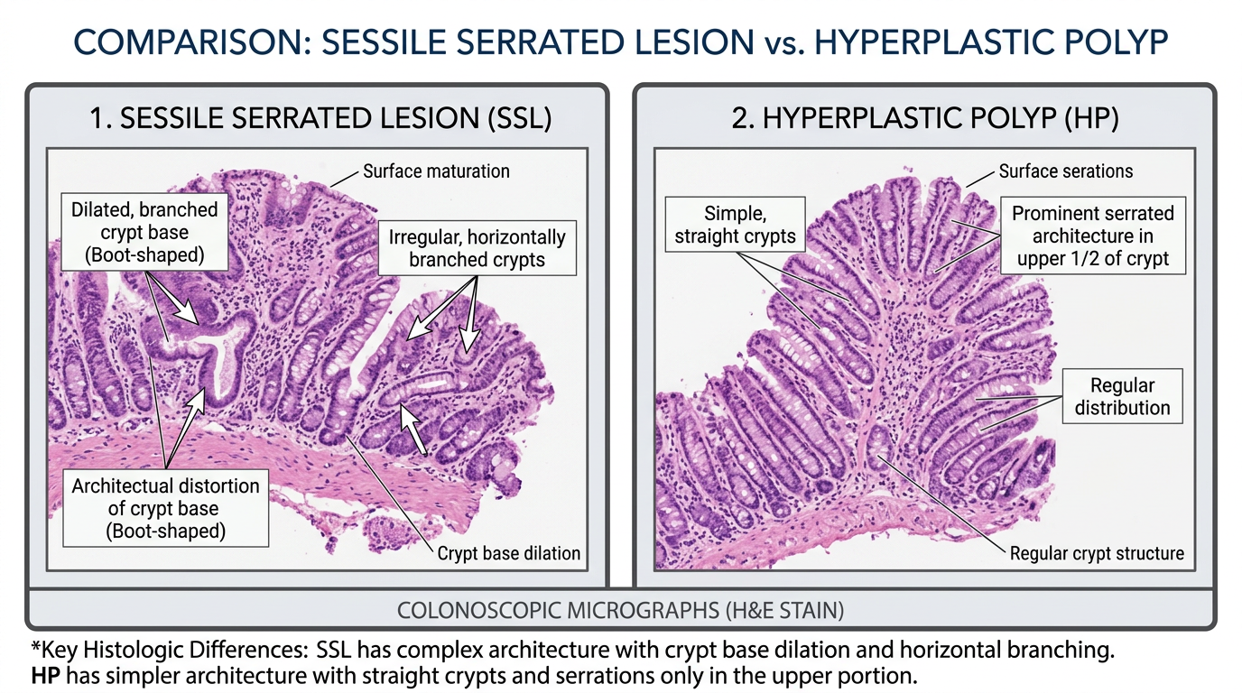
A pathology report may mention both sessile serrated lesion and hyperplastic polyp because these diagnoses can overlap in appearance, especially in small samples. The surface of both lesions may show a serrated polyp pattern, and the distinction often depends on deeper gland architecture. If the colonoscopy biopsy is fragmented or only samples the top of the polyp, the most diagnostic area may be limited. That is one reason the final diagnosis may include cautious language or a comment about clinical correlation.
Location in the colon also matters. A small hyperplastic polyp in the rectum or sigmoid colon is commonly low risk. A similar-looking serrated polyp in the right side of the colon may receive closer attention because right-sided lesions are more often part of the serrated pathway. The serrated pathway is one route by which some colon polyps can develop molecular changes over time and, in certain circumstances, contribute to colorectal cancer. This does not mean that every sessile serrated lesion becomes cancer, but it explains why correct classification matters.
Pathologists also look for dysplasia, which means the cells have developed more abnormal growth features. Dysplasia risk is not the same for every colon polyp, and a sessile serrated lesion without dysplasia is different from one with dysplasia. The presence of dysplasia can influence follow-up because it suggests the polyp has moved further along a precancerous sequence. Reports may also describe size, completeness of removal, and whether the specimen was received in pieces, all of which help the treating doctor plan surveillance.
How Serious Is a Sessile Serrated Lesion?
A sessile serrated lesion is usually serious enough to deserve proper follow-up, but it is not the same thing as a cancer diagnosis. Many are completely removed during colonoscopy and never cause harm. The concern is future risk, especially when the lesion is large, located in the right colon, incompletely removed, or shows dysplasia. In that setting, the report helps prevent colorectal cancer by identifying a polyp type that should not be ignored.
The difference from a hyperplastic polyp is mainly about risk category. A tiny left-sided hyperplastic polyp often carries very little concern and may not shorten the surveillance interval much. A sessile serrated lesion may lead to an earlier repeat colonoscopy because it is recognized as a precursor lesion in the serrated pathway. The exact interval depends on national guidelines, number of polyps, polyp size, dysplasia risk, quality of bowel preparation, and whether removal was complete.
Uncertainty can happen because a colonoscopy biopsy may be small, cauterized, or fragmented. Sometimes one pathologist may favor hyperplastic polyp, while another may favor sessile serrated lesion, especially in borderline cases. This is not a sign that anyone was careless; it reflects how subtle these gland patterns can be. When the diagnosis could change follow-up timing, a second review can be reasonable, and guidance on that process is available in When Should You Get a Second Pathology Opinion?.
What Happens Next: Treatment and Monitoring
The main treatment for a sessile serrated lesion is complete removal during colonoscopy when possible. If the report says the polyp was removed in fragments, was large, or had uncertain margins, the gastroenterologist may recommend a shorter interval before the next exam. A hyperplastic polyp may need less intensive monitoring, particularly when it is small and located in the distal colon. The colonoscopy biopsy result is therefore only one part of the full decision.
Follow-up also depends on how many polyps were found. Multiple serrated polyp findings may raise different questions than a single small lesion. The endoscopy report, bowel preparation quality, family history, and prior colonoscopy history all help determine the surveillance plan. A patient’s doctor may use these details to decide whether the next colonoscopy should occur in months, a few years, or a longer interval.
If dysplasia is reported, the conversation often becomes more focused. Dysplasia risk does not automatically mean cancer, but it means the cells show more advanced precancerous change. A sessile serrated lesion with dysplasia usually prompts careful confirmation of complete removal and closer surveillance. More general background about report structure is available in What Is a Pathology Report?.
Questions to Ask Your Doctor or Pathologist
- Was the polyp diagnosed as a sessile serrated lesion, a hyperplastic polyp, or another type of serrated polyp?
- Where in the colon was the colon polyp located?
- How large was the polyp, and was it removed completely?
- Did the colonoscopy biopsy show any dysplasia?
- Was the specimen fragmented or cauterized in a way that made interpretation harder?
- How does this result affect the timing of the next colonoscopy?
- Would a second pathology review change the follow-up recommendation?
These questions can help turn a confusing report into a practical plan. A sessile serrated lesion diagnosis is often most meaningful when combined with the colonoscopy findings, not read in isolation. A hyperplastic polyp in one location may have different implications than a serrated polyp in another location.
If wording in the pathology report feels unclear, an expert review may help confirm whether the diagnosis is best classified as sessile serrated lesion or hyperplastic polyp. That can be especially useful when surveillance timing depends on the distinction. Information about the review process is available at How to Get a Second Opinion on Your Pathology Diagnosis.
Frequently Asked Questions
Is a sessile serrated lesion cancer?
No, a sessile serrated lesion is not cancer by itself. It is considered a precancerous type of colon polyp because some lesions can contribute to colorectal cancer over time if they are not removed. Most are found and removed before cancer develops. The pathology report should state if invasive cancer is present, and that is a different diagnosis.
What is the difference between sessile serrated lesion and hyperplastic polyp?
The main difference is the microscopic architecture and the level of future risk. A hyperplastic polyp is often low risk, especially when small and located in the rectum or sigmoid colon. A sessile serrated lesion has deeper gland changes that can place it in the serrated pathway. That is why it may lead to closer colonoscopy follow-up.
Can a hyperplastic polyp be misdiagnosed?
Borderline cases can be difficult, especially when the colonoscopy biopsy is small, fragmented, or lacks the base of the glands. A hyperplastic polyp and a sessile serrated lesion can both show serrated surface changes. Pathologists rely on crypt architecture, location, and specimen quality to make the best diagnosis. A second opinion can be helpful when the distinction changes surveillance.
Does dysplasia in a serrated polyp mean cancer?
Dysplasia means the cells show more abnormal precancerous changes, but it does not automatically mean cancer. Dysplasia risk becomes more clinically relevant when it occurs in a sessile serrated lesion or a larger serrated polyp. The report should clarify whether dysplasia is low grade, high grade, or associated with invasive cancer. The treating physician uses that information to plan follow-up.
How often do I need colonoscopy after a sessile serrated lesion?
The interval depends on size, number of polyps, location, dysplasia, completeness of removal, and colonoscopy quality. A sessile serrated lesion may lead to earlier surveillance than a small distal hyperplastic polyp. The gastroenterologist should combine the pathology report with the colonoscopy report before giving a final interval. Patients can ask for the specific reason behind the recommended timing.
A report mentioning a sessile serrated lesion can be unsettling, but it often represents an opportunity for prevention rather than a crisis. The most useful next step is to clarify the exact diagnosis, removal status, and follow-up plan. Honest Pathology consultations can provide an independent pathology review when the wording is confusing or when a second opinion may affect surveillance decisions.
References:
National Cancer Institute — Pathology Reports Fact Sheet
National Cancer Institute — Definition of Polyp
NCBI Bookshelf — Medical Reference Library
