A celiac biopsy score can feel confusing. Marsh classification celiac biopsy results become clearer with this plain-English next-step guide.
Marsh classification celiac biopsy results describe how much the lining of the small intestine looks changed under the microscope in a pattern that can support celiac disease. The score is not a cancer stage, and it is not a measurement of how much pain or fatigue a patient feels. It is a structured way for a pathologist to describe inflammation and injury in tiny samples from the duodenum.
Seeing words like “Marsh 1,” “Marsh 3,” “villous atrophy,” or “intraepithelial lymphocytes” can make a pathology report feel more frightening than helpful. That reaction is understandable. A biopsy report uses compressed medical language, and even one line can carry a lot of meaning. The goal is to translate that line into a practical explanation that helps a patient prepare for the next medical conversation with less fear and more clarity.
Marsh Classification Celiac Biopsy — What It Actually Means
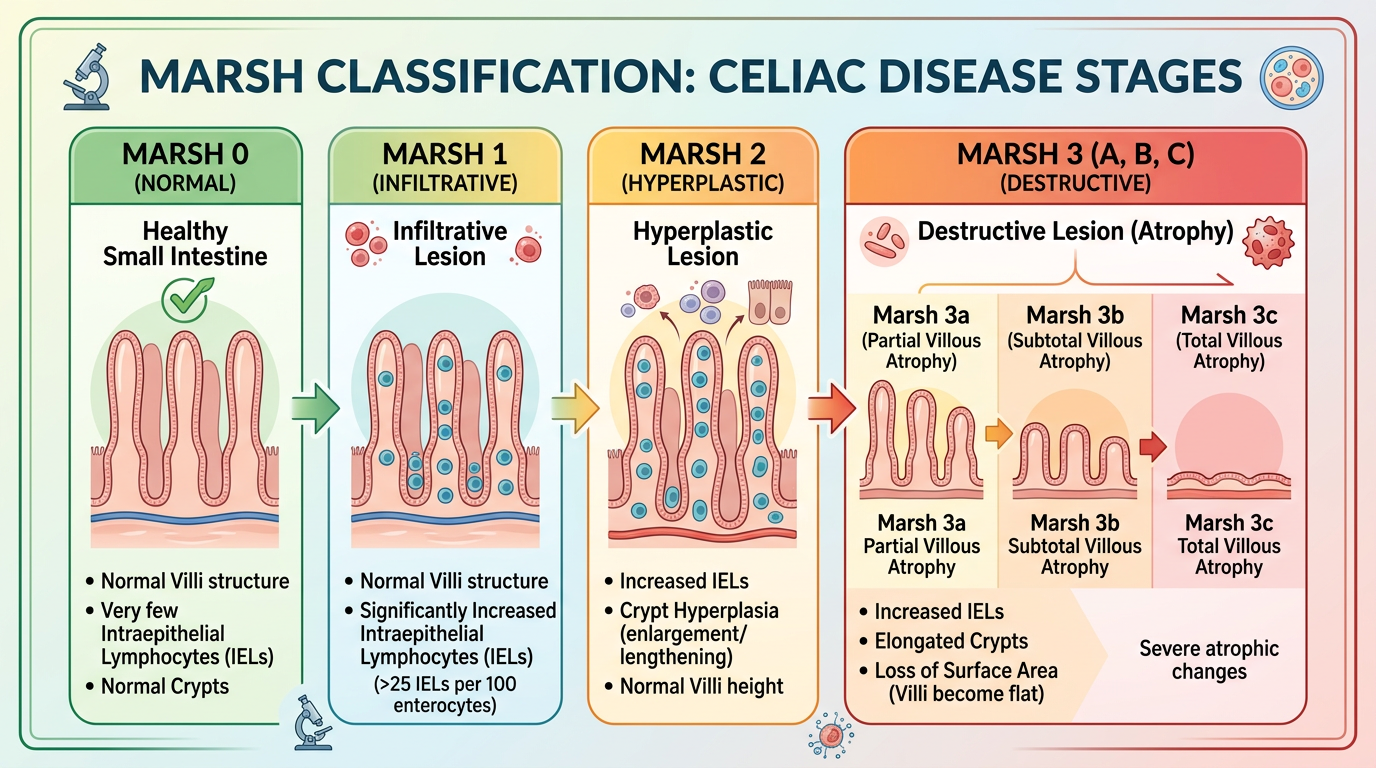
The Marsh classification celiac biopsy system is a microscopic scoring system used when a pathologist evaluates tissue from the duodenal mucosa, the first part of the small intestine beyond the stomach. In celiac disease, gluten exposure can trigger an immune reaction that injures this lining. The Marsh score describes a spectrum, from increased immune cells alone to more obvious flattening of the normal fingerlike projections called villi. A celiac disease biopsy is therefore not judged by one feature alone, but by a pattern of changes.
A helpful analogy is to picture the small intestine lining as a soft carpet with tall fibers. Healthy villi increase the surface area for absorbing nutrients, much like tall carpet fibers create a large surface. In more advanced celiac-type injury, those fibers become shortened or flattened, which is called villous atrophy. At the same time, the pathologist counts immune cells called intraepithelial lymphocytes that sit within the surface lining. The combination of these findings creates the Marsh category.
Different reports may use the original Marsh system, the modified Marsh system, or the Marsh-Oberhuber classification. The wording may vary, but the basic concept remains similar: The score summarizes inflammation, crypt changes, and villous atrophy. A Marsh classification celiac biopsy result helps gastroenterologists combine the microscopic findings with symptoms, blood tests, and response to a gluten free diet. For broader help with report language, patients may also find Understanding Your Pathology Report useful when preparing questions for a clinician.
Why Your Report Shows This Finding

A Marsh score appears when a duodenal biopsy is taken to evaluate suspected celiac disease, unexplained anemia, chronic diarrhea, weight loss, bloating, nutritional deficiencies, or related symptoms. The pathologist examines the duodenal mucosa to look for the architecture of the villi, the depth of crypts, and the number of intraepithelial lymphocytes. In many cases, the report also comments on whether the sample is well oriented, because poorly oriented tissue can make the villi look shorter than they really are. This is one reason biopsy interpretation is careful, detail-based work rather than a quick visual guess.
Marsh 0 usually means the biopsy looks essentially normal. Marsh 1 usually means increased intraepithelial lymphocytes without definite villous atrophy. Marsh 2 adds crypt hyperplasia, meaning the regenerative glands are more active or enlarged. Marsh 3 includes villous atrophy and is often subdivided into partial, subtotal, or total villous atrophy. A celiac disease biopsy with Marsh 3-type changes is more strongly supportive of active celiac disease when the clinical setting fits.
Blood tests matter because the biopsy is only one part of the diagnosis. Tissue transglutaminase IgA, total IgA level, endomysial antibody, and sometimes deamidated gliadin peptide testing help determine whether the immune pattern fits celiac disease. The small intestine can also be affected by medications, infections, immune disorders, Crohn disease, tropical sprue, and other less common conditions. That is why a Marsh classification celiac biopsy result should be interpreted with the patient’s diet history, medication list, and tissue transglutaminase results. A pathology report gives a microscopic pattern; the final diagnosis often comes from combining that pattern with the full clinical picture.
How Serious Is Marsh Classification Celiac Biopsy?
The seriousness of a Marsh classification celiac biopsy result depends on the score, the symptoms, the blood tests, and whether the patient was eating gluten before the biopsy. Marsh 0 or Marsh 1 can be mild, early, treated, patchy, or nonspecific, especially if tissue transglutaminase testing is negative. Marsh 3, especially with clear villous atrophy, usually carries more diagnostic weight for active celiac-type injury. Even then, the result should not be read as a cancer stage or a prediction of permanent damage.
Marsh 1 can be one of the most confusing categories because it shows increased intraepithelial lymphocytes without clear villous atrophy. This pattern can be seen in early celiac disease, partially treated celiac disease, Helicobacter pylori infection, some medications such as nonsteroidal anti-inflammatory drugs, autoimmune conditions, infections, or other inflammatory settings. A celiac disease biopsy in this category often requires careful clinical correlation rather than an instant label. The report may sound concerning, but Marsh 1 does not automatically mean lifelong celiac disease. It means the lining has more immune cells than expected and the reason needs to be worked out.
Marsh 3 is typically more specific when the right clinical setting is present. Villous atrophy can interfere with absorption of iron, folate, vitamin D, calcium, and other nutrients, so clinicians often check for deficiencies. The gluten free diet can allow the duodenal mucosa to heal over time in many patients with confirmed celiac disease, although healing speed varies by age, adherence, baseline injury, and other medical factors. A Marsh classification celiac biopsy result can therefore be serious enough to deserve follow-up, but it is also often actionable. The key is not panic; the key is confirming the cause and making a realistic treatment plan.
What Happens Next: Treatment and Monitoring
After a Marsh classification celiac biopsy result, the next step is usually a discussion with a gastroenterologist who can match the biopsy to blood work and symptoms. If celiac disease is confirmed, treatment usually centers on a strict gluten free diet guided by a knowledgeable clinician or dietitian. This diet is not a casual food preference in confirmed celiac disease; it is the medical treatment that removes the immune trigger. Follow-up may include tissue transglutaminase testing, nutritional labs, symptom review, and assessment for accidental gluten exposure.
Patients should not start or stop gluten before testing without medical guidance, because diet changes can alter both blood tests and biopsy findings. If a patient was already avoiding gluten before endoscopy, the biopsy may show less injury and the blood tests may be lower or negative. In some situations, a clinician may discuss a gluten challenge before repeat testing, but that decision depends on symptoms and risk. A celiac disease biopsy is most informative when the testing conditions are known and documented.
Repeat biopsy is not needed for every patient, but it may be considered when symptoms persist, blood tests remain abnormal, the original diagnosis is uncertain, or complications are suspected. Persistent villous atrophy despite a gluten free diet may lead clinicians to review diet exposure, medication effects, immune conditions, or rare refractory celiac disease. The original slides can also be reviewed if the diagnosis does not fit the clinical picture. For patients unsure whether another review is appropriate, When Should You Get a Second Pathology Opinion? explains common reasons to seek another pathology review.
Questions to Ask Your Doctor or Pathologist
Specific questions can turn a confusing biopsy report into a focused conversation. A Marsh classification celiac biopsy result should prompt questions about the score itself, the quality of the tissue sample, and how well the microscopic pattern matches the rest of the evaluation. Patients often benefit from bringing the pathology report, blood test results, medication list, diet history, and symptom timeline to the appointment. Clear questions can also help separate confirmed celiac disease from conditions that mimic it.
- What Marsh score was assigned, and was it original Marsh, modified Marsh, or Marsh-Oberhuber terminology?
- Did the biopsy show villous atrophy, increased intraepithelial lymphocytes, crypt hyperplasia, or all three?
- Was the duodenal mucosa well oriented enough for accurate assessment of villous height?
- Were enough duodenal biopsy samples taken from the bulb and distal duodenum?
- How do the biopsy findings compare with tissue transglutaminase and other celiac blood tests?
- Could medication use, infection, autoimmune disease, or another small intestine disorder explain the findings?
- Should the original slides be reviewed before committing to a lifelong gluten free diet?
A second pathology review can be especially helpful when the report shows borderline Marsh 1 findings, when the biopsy and blood tests do not match, or when treatment decisions feel uncertain. This does not mean the first report is wrong; it means that subtle small bowel biopsies can benefit from expert correlation.
Patients seeking another review can learn the practical steps through How to Get a Second Opinion on Your Pathology Diagnosis. For anyone new to biopsy terminology, What Is a Pathology Report? offers a clear foundation for understanding what a report can and cannot answer.
Frequently Asked Questions
What does Marsh 3 mean on a celiac biopsy?
Marsh 3 means the biopsy shows villous atrophy along with the inflammatory pattern often associated with active celiac disease. The villi are shortened or flattened to some degree, which can reduce nutrient absorption. The diagnosis is strongest when the biopsy matches positive celiac blood tests and symptoms. Marsh 3 is not a cancer stage, and it often improves with a strict gluten free diet when celiac disease is the cause.
Can Marsh 1 mean celiac disease?
Marsh 1 can be seen in celiac disease, but it is not specific by itself. It means there are increased intraepithelial lymphocytes without definite villous atrophy. A Marsh classification celiac biopsy with Marsh 1 findings needs correlation with tissue transglutaminase results, diet history, medications, and symptoms. Other conditions can cause the same microscopic pattern.
Can a celiac biopsy be wrong?
A biopsy can be limited by patchy disease, too few samples, poor tissue orientation, or gluten restriction before testing. The interpretation can also be challenging when changes are mild. A celiac disease biopsy is most reliable when multiple well-oriented samples are examined and the clinical information is available. If the result and the patient’s story do not fit, slide review by another pathologist may help.
Does villous atrophy always mean celiac disease?
Villous atrophy strongly raises the possibility of celiac disease, especially with positive celiac blood tests. However, villous atrophy can also occur with some infections, immune disorders, medication-related injury, and other small intestine diseases. A Marsh classification celiac biopsy result should therefore be interpreted in context. The cause matters because treatment depends on the underlying reason for the injury.
How long does it take for a gluten free diet to heal the intestine?
Healing varies from person to person. Symptoms may improve within weeks, while the duodenal mucosa can take months to years to recover fully, especially in adults. Follow-up blood tests such as tissue transglutaminase often help monitor response, although they do not show the lining directly. Persistent symptoms may require review for hidden gluten exposure, nutritional deficiencies, or another diagnosis.
A Marsh score is a microscope-based description, not a judgment on a patient’s future. With the right clinical correlation, most patients can move from uncertainty to a clear plan. Honest Pathology consultations can help patients and families understand whether the biopsy wording, score, and clinical picture fit together.
References:
National Cancer Institute — Pathology Reports
MedlinePlus — Celiac Disease
NCBI Bookshelf — Medical Reference Library