A kidney biopsy can feel frightening. lupus nephritis biopsy results class I–VI explain kidney damage and guide the next treatment questions.
lupus nephritis biopsy results divide kidney involvement from lupus into Classes I–VI, and each class describes a different pattern of kidney injury under the microscope. These classes are not random labels; they are a structured way for pathologists and kidney specialists to estimate activity, chronic damage, and treatment needs. A kidney biopsy result often becomes the turning point that helps a care team decide whether close monitoring, immune treatment, or long-term kidney protection is needed.
Receiving a report with Roman numerals, unfamiliar stains, and words such as “proliferative,” “membranous,” or “sclerosis” can feel overwhelming. Many patients fear that one number means the worst possible outcome, but lupus kidney disease is more nuanced than that. The class matters, yet so do kidney function, urine findings, blood pressure, symptoms, and whether the biopsy shows active inflammation or old scarring. Clear pathology interpretation can turn a frightening report into a practical map for the next conversation.
Lupus Nephritis Biopsy — What It Actually Means
A lupus nephritis biopsy is a microscopic examination of kidney tissue to see how systemic lupus erythematosus is affecting the filtering units of the kidney. These filtering units are called glomeruli, and they act like tiny strainers that help remove waste while keeping blood proteins inside the body. When lupus causes immune system proteins to collect in these filters, the result can be inflammation, leakage of protein into the urine, and sometimes scarring. The kidney biopsy result gives a direct look at this process rather than relying only on blood and urine tests.
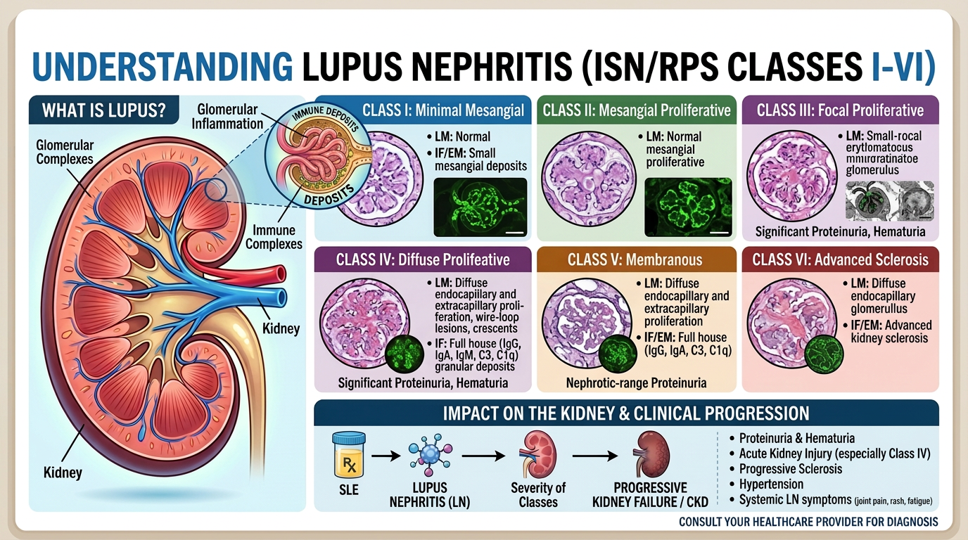
The International Society of Nephrology and Renal Pathology Society classification divides lupus nephritis into Classes I through VI. Class I and Class II are generally milder patterns, Class III and Class IV are proliferative patterns that often need stronger treatment, Class V is membranous lupus nephritis, and Class VI means advanced scarring. This classification is like a weather map for lupus kidney disease: It does not tell every detail of the future, but it shows where the storm is light, where it is active, and where damage may already be fixed. A lupus nephritis biopsy also helps separate active inflammation, which may improve with treatment, from chronic scarring, which is usually less reversible.
Pathologists often use light microscopy, immunofluorescence staining, and electron microscopy to classify the biopsy. Light microscopy shows the overall structure of the kidney filters and whether there is glomerular inflammation. Immunofluorescence staining highlights immune complex deposits, which are collections of antibodies and complement proteins that commonly appear in lupus kidney disease. Electron microscopy can show small immune complex deposits that are not always visible on routine stains, helping confirm the exact pattern behind the kidney biopsy result.
Why Your Report Shows This Finding

Lupus nephritis develops when the immune system forms antibodies that contribute to immune complex deposits in kidney tissue. These deposits can trigger complement activation and glomerular inflammation, leading to blood, protein, or cellular casts in the urine. A rising urine protein level may be one of the first clues that lupus kidney disease is active, even before a patient feels sick. Blood creatinine, estimated glomerular filtration rate, and urine sediment help clinicians decide when a biopsy is needed, but the biopsy shows the tissue-level pattern.
The class reported depends on where the immune complex deposits are located and how the kidney filters react to them. In Class I lupus nephritis, deposits may be seen mainly by immunofluorescence staining or electron microscopy, while the kidney tissue can look nearly normal under routine light microscopy. In Class II, there is mesangial involvement, meaning changes are mainly in the central support areas of the glomeruli. In Class III and Class IV, glomerular inflammation becomes more extensive and may involve segments of some glomeruli or many glomeruli throughout the biopsy.
Class V lupus nephritis has a membranous pattern, often associated with a higher urine protein level and swelling from protein loss. Class VI means most sampled glomeruli are globally sclerosed, or scarred, which suggests advanced chronic kidney injury rather than mainly active inflammation. A lupus nephritis biopsy report may also include an activity index and chronicity index, which help describe how much injury appears treatable versus permanent. Patients trying to understand the structure of a report may find it helpful to review what a pathology report includes before discussing the details with a nephrologist.
How Serious Is a Lupus Nephritis Biopsy Result?
The seriousness of a lupus nephritis biopsy result depends on the class, activity, chronicity, kidney function, and urine protein level. A lupus nephritis biopsy showing Class I or Class II may be reassuring when kidney function is stable and urine findings are mild. These classes still require clinical follow-up because lupus kidney disease can change over time, but they do not usually carry the same urgency as proliferative disease. The key question is not only “What class is it?” but also “Is the injury active, chronic, or both?”
Class III and Class IV are generally more serious because they represent focal or diffuse proliferative lupus nephritis. In these patterns, glomerular inflammation can damage kidney filters quickly if it is not treated effectively. The report may mention crescents, necrosis, endocapillary hypercellularity, or wire-loop changes, which are signs pathologists use to describe active injury. A lupus nephritis biopsy with high activity and low chronicity can sound alarming, but it may also mean there is inflammation that treatment can still control.
Class V can be serious in a different way because it may cause heavy protein loss, sometimes called nephrotic-range proteinuria. A very high urine protein level can lead to swelling, low blood albumin, and an increased risk of blood clots. Class VI is serious because it reflects extensive chronic scarring, and immune suppression may have limited benefit when most kidney filters are already replaced by scar tissue. Still, even advanced lupus kidney disease can have meaningful treatment goals, including blood pressure control, protein reduction, kidney protection, and planning ahead with the care team.
What Happens Next: Treatment and Monitoring
After a lupus nephritis biopsy, treatment is usually guided by a nephrologist and rheumatologist together. Class I and Class II may be managed with lupus control, blood pressure treatment, kidney-protective medications, and monitoring rather than aggressive immune suppression. Class III and Class IV commonly require immunosuppressive therapy because glomerular inflammation can progress without treatment. Class V may require treatment when the urine protein level is high, kidney function declines, or symptoms such as swelling are significant.
Monitoring typically includes urine protein level, urinalysis, serum creatinine, estimated glomerular filtration rate, complement levels, anti-dsDNA antibody levels, and blood pressure. These tests do not replace the kidney biopsy result, but they help show whether treatment is working over time. A falling urine protein level often suggests improvement, while persistent blood or protein in the urine may prompt treatment adjustment. In selected cases, a repeat lupus nephritis biopsy may be considered if the clinical picture changes or if treatment response is unclear.
Pathology reports can be difficult because they condense a large amount of microscopic information into short technical phrases. Reading the report with a structured approach can reduce fear and prevent a single scary word from taking over the entire interpretation. A patient-friendly explanation of report sections is available in Understanding Your Pathology Report: How to Read It with Confidence. When a kidney biopsy result will influence strong medication choices, a second expert review may also help confirm the class, activity, chronicity, and presence of immune complex deposits.
Questions to Ask Your Doctor or Pathologist
- What lupus nephritis class was found, and was more than one class present?
- Does the kidney biopsy result show mostly active inflammation, chronic scarring, or both?
- What are the activity index and chronicity index, if they were reported?
- How high is the urine protein level, and is it improving or worsening?
- Did immunofluorescence staining and electron microscopy support the same diagnosis?
- Were there crescents, necrosis, thrombotic microangiopathy, or other high-risk features?
- Would a second pathology opinion change treatment planning or provide added confidence?
These questions help turn a lupus nephritis biopsy report into a focused clinical discussion. The goal is not to memorize every microscopic term, but to understand which findings are driving treatment decisions. A clear conversation can also help separate urgent findings from findings that simply need careful monitoring.
A second opinion is most useful when the class is unclear, the biopsy sample is limited, the findings do not match the clinical picture, or major immune suppression is being considered. Helpful background on when review may be appropriate is available in When Should You Get a Second Pathology Opinion?. When needed, families can also learn the practical steps in How to Get a Second Opinion on Your Pathology Diagnosis.
Frequently Asked Questions
What does lupus nephritis class mean?
A lupus nephritis class describes the pattern of kidney injury seen under the microscope. Classes I through VI are based on where immune complex deposits are located, how much glomerular inflammation is present, and whether scarring has developed. The class helps clinicians estimate risk and choose treatment. It should always be interpreted with kidney function, urine protein level, and the patient’s overall lupus activity.
Is class 4 lupus nephritis the worst?
Class IV is often considered one of the more serious active forms because it involves many glomeruli and can progress without treatment. However, “worst” is not always the right word because active Class IV disease may respond to therapy, especially when chronic scarring is limited. A lupus nephritis biopsy with Class IV and a high activity index can be urgent, but it may still represent treatable inflammation. Class VI may be more concerning for permanent damage because it reflects advanced scarring.
Can lupus nephritis biopsy results change over time?
Yes, lupus kidney disease can evolve, and a later biopsy may show a different class from the first biopsy. Treatment can reduce active inflammation, while untreated or severe disease can leave chronic scarring. A repeat lupus nephritis biopsy may be considered when urine findings worsen, kidney function changes, or the clinical course does not match the original kidney biopsy result. Decisions about repeat biopsy are individualized.
What is the difference between activity and chronicity?
Activity refers to inflammation or injury that may improve with treatment, such as cellular proliferation, necrosis, or crescents. Chronicity refers to scarring or permanent structural damage, such as global sclerosis, fibrous crescents, tubular atrophy, or interstitial fibrosis. A kidney biopsy result with high activity and low chronicity may justify stronger treatment because there is potentially reversible injury. High chronicity suggests that kidney-protective care and long-term planning become especially important.
Should I get a second opinion on a lupus nephritis biopsy?
A second opinion can be helpful when the biopsy class is uncertain, the sample is small, or treatment decisions carry major risks. Renal pathology is specialized, and review by a pathologist experienced in kidney disease can confirm findings such as immune complex deposits, glomerular inflammation, and chronic scarring. A lupus nephritis biopsy second review does not mean the first report was wrong; it means the diagnosis is being checked before important decisions are made. This can provide clarity for patients, caregivers, and treating physicians.
A lupus nephritis biopsy result can feel like a verdict, but it is better understood as a detailed map of kidney injury. The most helpful next step is a careful explanation of the class, the activity, the chronicity, and how those findings match the urine and blood tests. Honest Pathology consultations can help patients and families understand the pathology language before treatment decisions are finalized.
References:
National Cancer Institute — Pathology Reports Fact Sheet
MedlinePlus — Lupus
NCBI Bookshelf — Biomedical And Clinical Reference Library