

Worried by indefinite for dysplasia in Barrett’s esophagus? Learn what this biopsy result means and what to ask next.
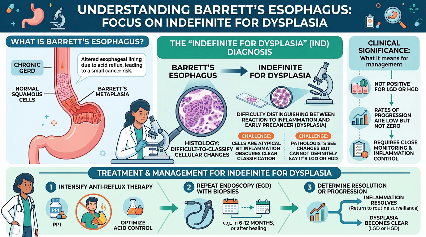
indefinite for dysplasia means the biopsy has abnormal-looking cells, but the pathologist cannot confidently say whether the changes are truly precancerous or caused by irritation and inflammation.
In Barrett’s esophagus, this wording often appears when a Barrett’s esophagus biopsy shows intestinal metaplasia with cellular changes that sit in a gray zone. The result can feel unsettling because the word “dysplasia” is present, yet the report does not give a clear yes-or-no answer. Pathologists use this category carefully because treatment decisions in Barrett’s esophagus depend heavily on whether true esophageal dysplasia is present.
Receiving this result can make a patient feel as if the ground has shifted. The most helpful first step is to understand that indefinite for dysplasia is not the same as cancer, and it is not the same as definite high-grade dysplasia. It is a signal that the tissue needs careful correlation, acid control, and often expert review.
A pathology report can be difficult to read even when the result is straightforward. A patient who needs the basics may benefit from reviewing what a pathology report is before focusing on the Barrett’s-specific language.
Indefinite For Dysplasia — What It Actually Means
Indefinite for dysplasia is a diagnostic category used when the lining cells in Barrett’s esophagus look more abnormal than expected, but the changes are not convincing enough to diagnose definite esophageal dysplasia. In simple terms, the biopsy looks suspicious, but not proven. It is like seeing smoke near a kitchen: There may be a fire, or there may only be steam from a hot pan. The pathologist is acknowledging concern while avoiding an overdiagnosis that could lead to unnecessary procedures.
In a Barrett’s esophagus biopsy, the pathologist examines tiny pieces of tissue under the microscope. Barrett’s esophagus means that the normal squamous lining of the lower esophagus has been replaced by intestinal metaplasia, a gland-forming lining that resembles tissue from the intestine. This change usually develops after long-term acid reflux inflammation. Once intestinal metaplasia is present, the biopsy is evaluated for dysplasia, which means precancerous change in the cells.
The challenge is that acid reflux inflammation can make cells look enlarged, crowded, and reactive. These reactive changes can overlap with early esophageal dysplasia. The term indefinite for dysplasia exists because biology is sometimes messy, especially in inflamed tissue. A careful report gives the treating gastroenterologist enough information to improve acid control, repeat sampling if needed, and consider a pathology second opinion when the features are borderline.
Why Your Report Shows This Finding
A report may show indefinite for dysplasia because the biopsy contains both Barrett’s-type tissue and active injury. Acid reflux inflammation can damage the surface lining, cause erosions, and trigger repair. During repair, glandular cells may become enlarged, darker, and more crowded than usual. This pattern is called glandular atypia, and it can be reactive rather than precancerous.
Pathologists look at several microscopic clues before calling definite esophageal dysplasia. They assess how crowded the glands are, whether the nuclei are enlarged and elongated, whether the cells have lost their normal maturation pattern, and whether the abnormality is present away from inflamed surface injury. In indefinite for dysplasia, some of those features are present, but the overall pattern is not strong enough for a definitive diagnosis. The biopsy may also be small, fragmented, cauterized, or oriented in a way that makes interpretation harder.
Medication history also matters. A proton pump inhibitor may reduce acid injury and make a repeat Barrett’s esophagus biopsy easier to interpret. When inflammation is active, the pathologist may see glandular atypia that would look less worrisome after healing. For that reason, doctors often focus first on controlling acid reflux inflammation, then reassessing the lining after treatment. This approach helps separate true esophageal dysplasia from changes caused by injury and repair.
How Serious Is Indefinite For Dysplasia?
Indefinite for dysplasia is serious enough to deserve follow-up, but it should not be understood as a cancer diagnosis. The finding means the biopsy falls between negative for dysplasia and definite dysplasia. Some patients later have no dysplasia on repeat biopsy after acid reflux inflammation improves. Others may later be found to have low-grade or, less commonly, high-grade esophageal dysplasia, especially if the original biopsy had concerning features.
The seriousness depends on several details in the report and clinical situation. A single tiny area of glandular atypia in a very inflamed Barrett’s esophagus biopsy may be less concerning than repeated indefinite for dysplasia diagnoses across multiple biopsy levels. A long segment of Barrett’s esophagus, visible nodules, ulcers, or prior dysplasia may raise the level of concern. The pathologist’s comments, the endoscopy findings, and the number and location of biopsies all help place indefinite for dysplasia in context.
In real-world practice, the main danger is misunderstanding the category. If indefinite for dysplasia is dismissed completely, a patient may miss needed surveillance. If it is treated as definite high-grade dysplasia, a patient may feel unnecessary fear and may undergo more aggressive intervention than the evidence supports. The balanced response is careful acid suppression, repeat endoscopy at an appropriate interval, and consideration of a pathology second opinion when the result will affect management.
A patient trying to read the entire report may also find this patient-focused explanation of how to read a pathology report helpful. The wording around dysplasia, margins, fragments, and comments often becomes clearer when the report is broken into its main parts.
What Happens Next: Treatment And Monitoring
After an indefinite for dysplasia result, many gastroenterologists first intensify acid suppression. A proton pump inhibitor is commonly used because reducing reflux injury can allow the lining to heal. The goal is not to “treat dysplasia” with acid medicine alone, but to remove inflammation that may be mimicking dysplasia. Once acid reflux inflammation improves, the next Barrett’s esophagus biopsy may be more reliable.
Repeat endoscopy is often recommended, although timing depends on the patient’s history, endoscopic findings, and local guidelines. During repeat endoscopy, the doctor may take systematic biopsies throughout the Barrett’s segment and target any visible abnormal areas. If esophageal dysplasia is confirmed, management may change to closer surveillance or endoscopic therapy. If the repeat biopsy is negative for dysplasia, the patient may return to a surveillance plan based on the Barrett’s segment length and risk profile.
A pathology second opinion can be especially helpful when the diagnosis is indefinite for dysplasia, low-grade dysplasia, or high-grade dysplasia. These categories can be difficult, and even experienced pathologists may request another gastrointestinal pathology review. A second review does not mean the first pathologist made a mistake. It means the diagnosis has enough clinical weight that confirmation is often wise before major treatment decisions.
Questions To Ask Your Doctor Or Pathologist
- Was the biopsy diagnosis indefinite for dysplasia, low-grade dysplasia, or high-grade dysplasia?
- Was active acid reflux inflammation present in the Barrett’s esophagus biopsy?
- Did the report describe intestinal metaplasia, glandular atypia, or any visible lesion that was biopsied?
- Should acid suppression with a proton pump inhibitor be increased before repeat endoscopy?
- How long is the Barrett’s segment, and were biopsies taken from the full area?
- Should the slides be reviewed by a gastrointestinal pathologist through a pathology second opinion?
- What follow-up interval is recommended if the next biopsy is negative for esophageal dysplasia?
These questions help turn an unclear report into a practical plan. The goal is not to memorize pathology terminology, but to understand whether the result reflects true precancerous change, inflammation-related mimicry, or a finding that needs confirmation.
When a report contains borderline language, expert review can be reassuring and clinically useful. More information about when a review may matter is available in this guide to when to get a second pathology opinion. For patients already considering slide review, this resource explains how to get a second opinion on a pathology diagnosis.
Frequently Asked Questions
Is indefinite for dysplasia cancer?
No. Indefinite for dysplasia is not cancer. It means the cells look abnormal, but the pathologist cannot confidently classify them as definite precancerous dysplasia because inflammation or injury may be causing similar changes. Follow-up matters because some patients may later have definite esophageal dysplasia on repeat biopsy.
Can indefinite for dysplasia go away?
Yes, the finding can disappear on repeat biopsy, especially when acid reflux inflammation was strong in the original sample. Better acid control with a proton pump inhibitor may reduce reactive changes and make the next biopsy clearer. If the repeat Barrett’s esophagus biopsy shows no dysplasia, the doctor may adjust surveillance based on standard Barrett’s risk factors.
What causes indefinite for dysplasia in Barrett’s esophagus?
Common causes include reactive glandular atypia from reflux injury, erosions, ulceration, and repair in Barrett’s tissue. The biopsy may also be small or difficult to orient, making interpretation more limited. Indefinite for dysplasia is used when the pathologist sees concern but not enough evidence for definite esophageal dysplasia.
Should I get a second opinion for indefinite for dysplasia?
A pathology second opinion is often reasonable for indefinite for dysplasia, particularly if treatment decisions or follow-up timing depend on the result. Barrett’s dysplasia categories can be subtle, and review by a gastrointestinal pathologist may clarify the diagnosis. The review typically uses the original glass slides, so another endoscopy is not always needed for the second opinion itself.
How often does indefinite for dysplasia become high-grade dysplasia?
The risk varies based on the patient’s Barrett’s length, endoscopy findings, inflammation level, and whether the diagnosis persists on repeat biopsy. Many patients do not progress, especially when the original changes were driven by acid reflux inflammation. Persistent indefinite for dysplasia or confirmed esophageal dysplasia usually leads to closer surveillance and more specialized management.
Indefinite for dysplasia can sound frightening, but it is best understood as a careful warning rather than a final diagnosis. The next steps usually involve healing inflammation, repeating evaluation, and confirming the interpretation when needed. Honest Pathology consultations can help patients and caregivers understand the wording, the level of concern, and the questions to bring back to the treating team.
References:
National Cancer Institute — Pathology Reports
National Cancer Institute — Definition of Dysplasia
NCBI Bookshelf — Medical Texts and Clinical Reviews
