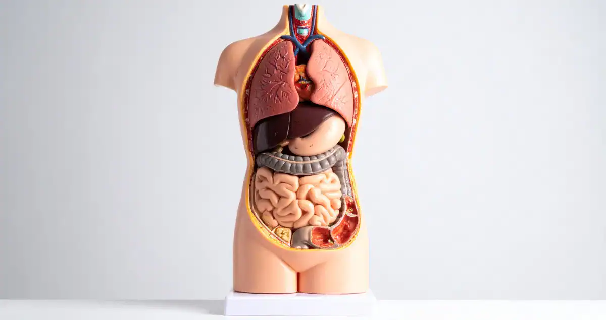
Biopsies of the kidney and liver are important tools for diagnosing a wide range of conditions. They provide critical information about how these organs are functioning, the presence of inflammation, scarring, infection, or other abnormalities, and guide treatment decisions. While lab tests and imaging studies offer clues about organ health, biopsy results allow pathologists to examine tissue under a microscope and provide a detailed understanding of disease processes. Learning how pathologists interpret and explain these results can help patients feel informed and confident about their care.
Why Kidney and Liver Biopsies Are Performed
Kidney and liver biopsies are typically recommended when routine blood tests, urine analysis, or imaging indicate abnormalities that require further clarification. For the kidney, this may include elevated creatinine, protein in the urine, or unexplained swelling. Liver biopsies may be suggested for elevated liver enzymes, persistent jaundice, or unexplained changes seen on imaging. Biopsies allow pathologists to look directly at tissue architecture, cell health, and signs of inflammation or scarring.
Pathologists are trained to interpret subtle changes in tissue structure and cellular detail, providing insights that cannot be obtained from blood tests alone. Honest Pathology principles emphasize clear, transparent explanations so patients understand both the findings and their significance.
How Biopsy Samples Are Processed
After the biopsy is collected, the tissue is sent to the laboratory for processing. It is fixed, embedded in paraffin, sliced into extremely thin sections, and stained with special dyes to highlight different tissue components. In kidney biopsies, stains may differentiate glomeruli, tubules, and blood vessels. Liver biopsies often use stains to show fibrosis, inflammation, fat, or iron deposits. In some cases, additional techniques such as immunofluorescence, immunohistochemistry, or electron microscopy are used to provide even more detailed information.
This meticulous processing allows pathologists to identify changes at both the microscopic and cellular level. For example, a kidney biopsy may reveal minimal change disease, focal segmental glomerulosclerosis, or immune complex deposition, each of which has distinct treatment implications. Liver biopsies may show fatty liver disease, autoimmune hepatitis, viral hepatitis, or cirrhosis, providing critical guidance for management.
The Structure of a Biopsy Report
Kidney and liver biopsy reports are typically divided into several sections. The gross description details the tissue’s appearance before microscopic examination, including size, color, and texture. The microscopic description explains what pathologists see under the microscope, such as the condition of glomeruli in the kidney or hepatocytes in the liver. Special stains and any additional studies are also described. The final diagnosis summarizes the findings and often includes a comment section providing context or recommendations for follow-up.
For instance, a kidney biopsy report might state, “Light microscopy shows focal segmental glomerulosclerosis with mild tubular atrophy. Immunofluorescence is negative. Electron microscopy reveals foot process effacement. Diagnosis: focal segmental glomerulosclerosis, tip variant.” Each of these details helps the treating physician understand the type and severity of the disease and plan appropriate therapy.

How Pathologists Explain Findings to Physicians
Pathologists communicate their findings to the treating physicians in a structured, precise manner. They describe the tissue architecture, the presence or absence of inflammation, fibrosis, or immune deposits, and any abnormal cellular features. They may provide differential diagnoses when findings are not entirely specific and suggest additional testing if needed.
For kidney biopsies, pathologists often focus on the glomeruli, tubules, interstitium, and blood vessels, noting changes such as immune complex deposition, inflammation, or scarring. In liver biopsies, they evaluate hepatocyte health, the degree of inflammation, fibrosis staging, fat accumulation, iron or copper deposition, and signs of bile duct injury.
Honest Pathology emphasizes that reports are written in a way that both physicians and patients can understand. Clear explanations about what is normal, what is abnormal, and what these findings mean for treatment help reduce confusion and anxiety.
Common Findings in Kidney Biopsies
Kidney biopsies reveal a wide variety of conditions. Minimal change disease, common in children, may show nearly normal glomeruli under light microscopy but subtle changes under electron microscopy. Focal segmental glomerulosclerosis presents with scarring in parts of some glomeruli, often causing proteinuria. Immune complex diseases like lupus nephritis reveal deposits that stain with immunofluorescence. Interstitial nephritis shows inflammation of the spaces between tubules, often due to medications or infection.
Pathologists explain how each finding affects kidney function and may influence treatment decisions. For example, the presence of scarring may indicate a more chronic process requiring long-term management, while minimal change disease may respond well to corticosteroids.
Common Findings in Liver Biopsies
Liver biopsies can uncover fatty liver disease, viral hepatitis, autoimmune hepatitis, biliary obstruction, or fibrosis leading to cirrhosis. Pathologists assess hepatocytes for degeneration or necrosis, inflammation patterns, bile duct changes, fat accumulation, and fibrosis staging. Special stains may highlight iron overload or copper deposition.
For instance, a report describing “mild portal and lobular inflammation with steatosis involving 20% of hepatocytes, no significant fibrosis” suggests early fatty liver disease, which may be managed with lifestyle changes. More advanced fibrosis or cirrhosis changes management significantly and may warrant closer monitoring or treatment interventions. Honest Pathology principles encourage explaining these nuances in ways patients can understand while still conveying the seriousness of advanced findings.
How Patients Can Understand Their Reports
Although biopsy reports are primarily written for physicians, patients can learn to read them with guidance. Key areas to focus on include the final diagnosis, description of the main abnormalities, and any comments about severity or prognosis. For example, a kidney biopsy noting “mild tubular atrophy” indicates some chronic damage but may not necessarily affect immediate treatment decisions. A liver biopsy stating “stage 3 fibrosis” reflects advanced scarring and may influence treatment and monitoring plans.
Patients should also understand that not all findings are definitive on their own. Biopsy interpretation is integrated with clinical data, lab results, and imaging to guide overall management. Honest Pathology supports transparent communication, helping patients grasp the meaning of complex terminology and microscopic details.
Questions Patients Might Consider Asking
During follow-up, patients may want to ask questions to clarify the significance of biopsy findings. Examples include asking how the findings affect kidney or liver function, what the likely progression of the condition is, whether additional testing is needed, and what treatment options are appropriate. For instance, a patient might ask whether mild inflammation in a liver biopsy requires medication or can be managed with lifestyle changes, or whether scarring in a kidney biopsy is reversible or permanent. Clear answers from healthcare providers help patients make informed decisions and reduce anxiety.
The Role of Follow-Up and Monitoring
Kidney and liver biopsy results often guide ongoing monitoring. Patients may have periodic blood tests, urine analysis, imaging, or repeat biopsies to track disease progression or response to treatment. Understanding the implications of biopsy findings helps patients engage in follow-up care. For example, fibrosis staging in liver biopsies often determines the frequency of monitoring and interventions to prevent further progression.
Conclusion
Pathologists play a critical role in explaining kidney and liver biopsy results. By examining tissue under the microscope, using special stains, and applying advanced techniques such as immunofluorescence or electron microscopy, they provide detailed insights into organ health, inflammation, scarring, and disease processes. Reports describe findings in structured language, highlighting both abnormalities and normal structures, and offer context for treatment planning. Honest Pathology principles encourage clear, transparent explanations that empower patients to understand their results, ask informed questions, and participate actively in their care. By combining biopsy data with clinical information, pathologists help create a complete picture of kidney and liver health, guiding personalized treatment decisions and follow-up strategies.