Scared by kidney biopsy results? FSGS minimal change disease can be confusing. Learn what the biopsy shows and what to ask next.
FSGS minimal change disease is a kidney biopsy distinction that helps explain why a patient may have heavy protein loss in the urine, swelling, and abnormal kidney tests. Both conditions can cause nephrotic syndrome, but they do not always behave the same way or respond to treatment in the same pattern. A kidney biopsy gives the treating team a closer look at the kidney filters, much like examining the wiring behind a flickering light rather than only seeing the bulb dim.
Receiving a report with terms such as focal segmental glomerulosclerosis, minimal change disease, podocyte injury, or electron microscopy can feel overwhelming. Many patients hear the word biopsy and immediately worry that the result must be catastrophic. In reality, the purpose of the biopsy is to reduce uncertainty, guide treatment, and help separate conditions that can look very similar from the outside.
This article explains what pathologists look for, why the distinction matters, and when a second review may help clarify the diagnosis.
FSGS Minimal Change Disease — What It Actually Means
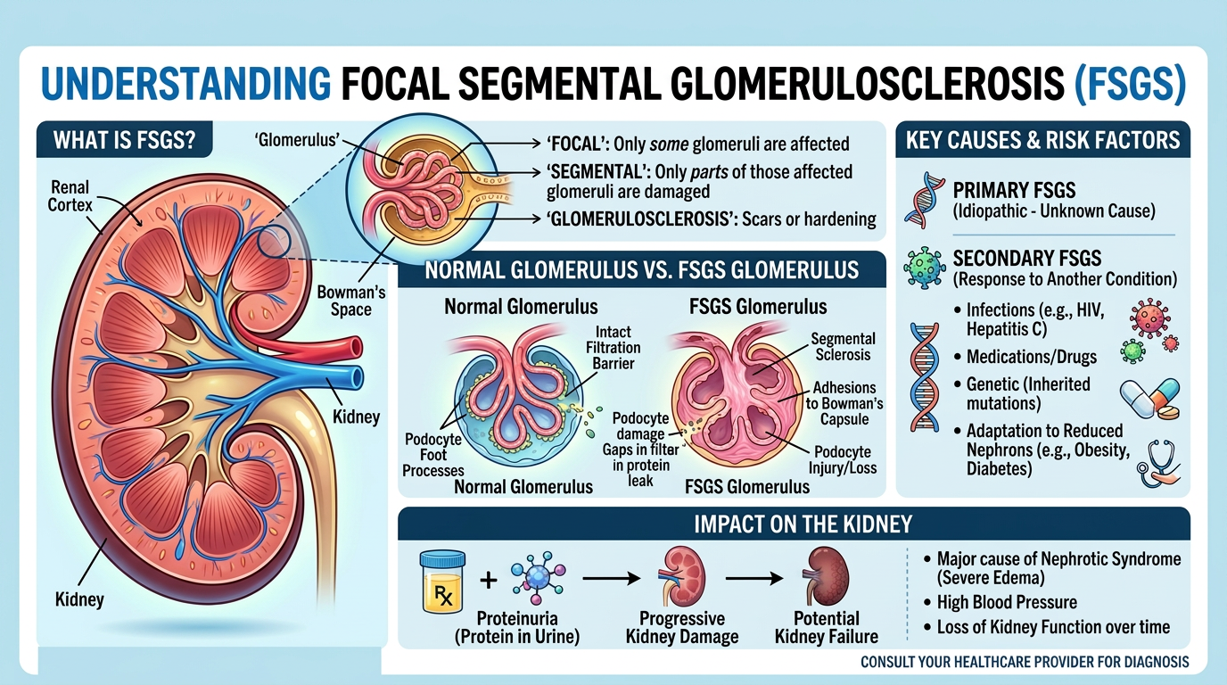
FSGS minimal change disease refers to two kidney biopsy patterns that can both cause major protein urine but show different structural clues under the microscope. Minimal change disease often means the kidney tissue looks nearly normal on routine light microscopy, even though the filtration cells are injured. FSGS stands for focal segmental glomerulosclerosis, which means some, but not all, kidney filtering units have scarring in part of the filter. That partial scarring is called segmental sclerosis, and it is one of the central clues pathologists search for during a kidney biopsy.
A helpful analogy is a row of coffee filters. In minimal change disease, the filter may look almost normal from a distance, but the tiny pores have become leaky. In FSGS, some filters show visible scarred patches that suggest a more established injury pattern. Both problems can lead to nephrotic syndrome because albumin and other proteins escape from the blood into the urine. This is why protein urine can be severe even when the kidney tissue does not look dramatically abnormal at first glance.
Pathologists evaluate several types of information before separating these diagnoses. Routine stains show the architecture of the glomeruli, immunofluorescence staining checks for immune deposits, and electron microscopy examines the very fine structures of the filtration barrier. The combination matters because minimal change disease and FSGS minimal change disease overlap in symptoms but not always in prognosis. For patients trying to understand the wording of a report, resources such as What Is a Pathology Report? can make the format less intimidating.
Why The Report Shows This Finding
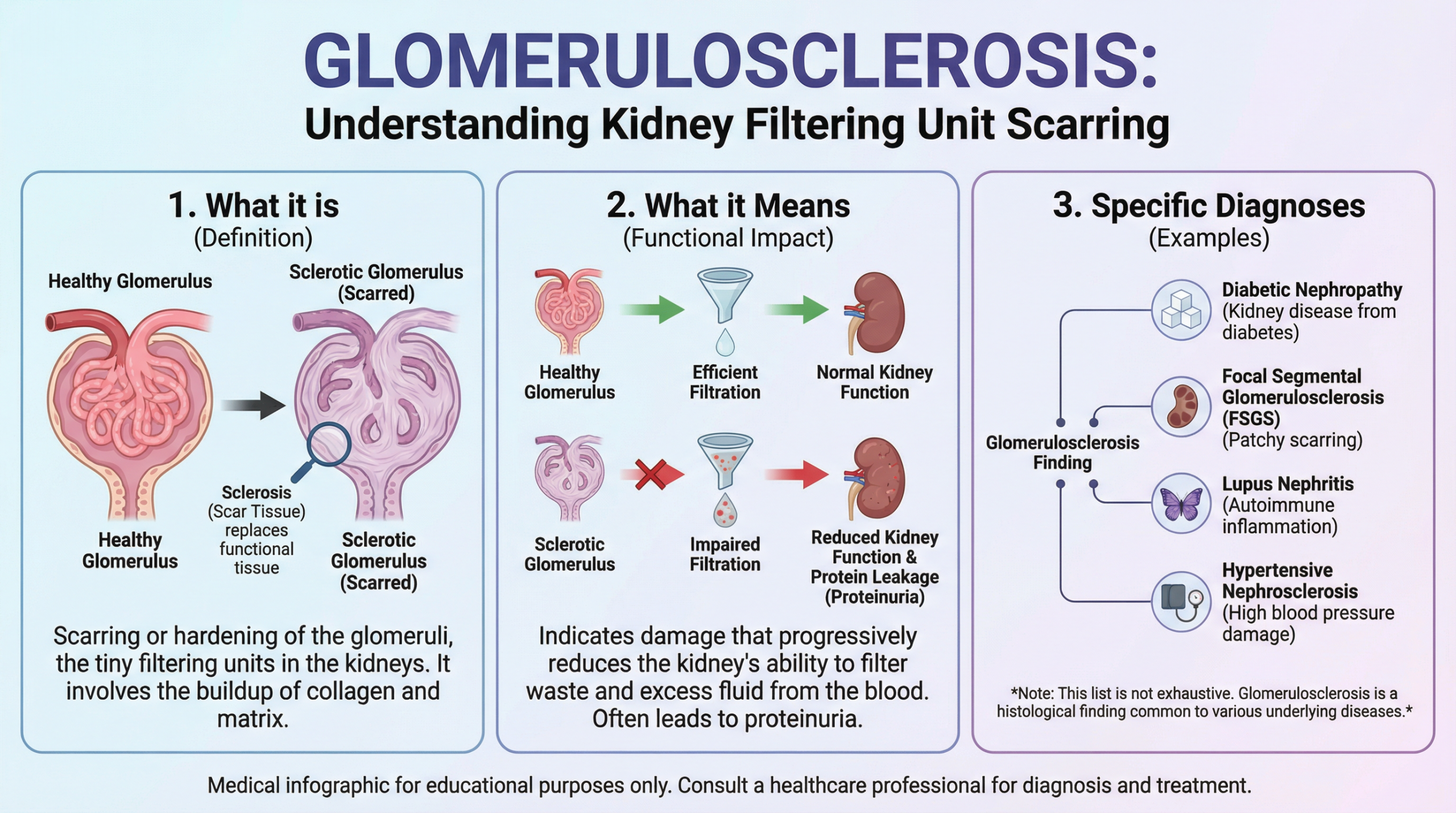
A kidney biopsy may show FSGS minimal change disease because the kidney filters have been injured at the level of the podocyte, a specialized cell that helps keep protein inside the bloodstream. Podocyte injury is a major concept in both diagnoses. When podocytes lose their delicate foot processes, the filtration barrier becomes leaky and protein urine increases. Minimal change disease is often dominated by this ultrastructural change, while FSGS also shows areas of segmental sclerosis on light microscopy.
The report may describe the number of glomeruli examined, how many are scarred, whether the scarring is global or segmental, and whether there are chronic changes in the surrounding kidney tissue. These details help the nephrologist estimate how active or longstanding the process appears. Immunofluorescence staining is used to look for immune complex diseases that can mimic or contribute to protein loss. If immunofluorescence staining is negative or nonspecific, that finding may support minimal change disease or a primary podocyte process, depending on the rest of the biopsy.
Electron microscopy is especially important because it can show diffuse foot process effacement, a hallmark of podocyte injury. In minimal change disease, electron microscopy may be the main place where the abnormality is clearly visible. In FSGS, electron microscopy may also show foot process effacement, but the light microscopy evidence of segmental sclerosis shifts the interpretation. Nephrotic syndrome can therefore look similar in the clinic while the biopsy reveals whether the pattern is more consistent with minimal change disease, FSGS, or a transition between the two.
How Serious Is FSGS Minimal Change Disease?
The seriousness of FSGS minimal change disease depends on the biopsy pattern, kidney function, amount of protein urine, response to treatment, and whether chronic scarring is already present. Minimal change disease often responds well to corticosteroid therapy, especially in children, though adults may respond more slowly. FSGS can be more variable, and some forms are harder to control. The diagnosis is not simply a label; it is a treatment map that helps the care team judge risk and choose the next step.
FSGS minimal change disease can be confusing because some biopsies show features that seem close to both categories. A biopsy sample is only a small portion of the kidney, and FSGS can be patchy. If the needle sample misses a scarred glomerulus, early FSGS may look like minimal change disease, especially when electron microscopy shows widespread foot process effacement. This sampling issue is one reason the number of glomeruli in the kidney biopsy report matters.
Several findings make the situation more concerning. Extensive segmental sclerosis, tubular atrophy, interstitial fibrosis, reduced kidney function, persistent nephrotic syndrome, and ongoing heavy protein urine can suggest a higher risk of long-term kidney damage. On the other hand, a biopsy with little chronic scarring, preserved kidney function, and a strong response to treatment is generally more reassuring. FSGS minimal change disease should be interpreted by combining pathology, blood tests, urine results, blood pressure, medication history, and clinical context.
What Happens Next: Treatment And Monitoring
After a diagnosis involving FSGS minimal change disease, the nephrologist typically reviews the biopsy findings alongside blood creatinine, albumin level, cholesterol, urine protein measurement, and blood pressure. Treatment may include corticosteroids, other immune-modifying medications, diuretics for swelling, and medications that reduce protein urine. The exact plan depends on whether the pattern looks like minimal change disease, primary FSGS, secondary FSGS, or another related podocyte disorder. Secondary FSGS can be associated with conditions such as obesity, reduced kidney mass, reflux nephropathy, certain medications, infections, or genetic factors.
Monitoring usually focuses on whether protein urine decreases and whether kidney function stabilizes. A falling urine protein level often provides an encouraging sign that the filtration barrier is healing. Persistent nephrotic syndrome may lead the doctor to adjust treatment or reconsider the diagnosis. In some cases, the original kidney biopsy may be reviewed again, especially if the clinical course does not match the expected pattern.
A pathology report can be dense, so structured reading can help. The article Understanding Your Pathology Report: How to Read It with Confidence explains how to approach unfamiliar terminology without getting lost in every phrase. When biopsy findings and symptoms do not seem to fit, a second review may be reasonable. Guidance on timing is available in When Should You Get a Second Pathology Opinion?.
Questions To Ask A Doctor Or Pathologist
- How many glomeruli were present in the kidney biopsy sample?
- Did the biopsy show definite segmental sclerosis, or was FSGS only suspected?
- What did electron microscopy show about foot process effacement?
- Was immunofluorescence staining negative, nonspecific, or suggestive of another disease?
- Does the pattern fit minimal change disease, primary FSGS, secondary FSGS, or an overlap pattern?
- How much chronic scarring, tubular atrophy, or interstitial fibrosis was present?
- How will protein urine be measured during treatment follow-up?
These questions help turn a technical report into a practical conversation. They also help separate what is known from what remains uncertain. A diagnosis involving FSGS minimal change disease often requires close communication between nephrology and pathology because the microscope findings must match the clinical picture.
A second pathology opinion can be helpful when the report uses uncertain wording, the biopsy sample is limited, or the treatment response does not fit the diagnosis. The process usually involves sending slides, special stains, electron microscopy images, and the original report for expert review. Practical steps are outlined in How to Get a Second Opinion on Your Pathology Diagnosis.
Frequently Asked Questions
Can minimal change disease turn into FSGS?
Minimal change disease does not simply transform in every patient, but the relationship can be complex. Some patients initially appear to have minimal change disease because the kidney biopsy sample does not capture a scarred glomerulus. Later findings or treatment response may support FSGS minimal change disease overlap or an early FSGS pattern. This is why electron microscopy, clinical follow-up, and protein urine trends are all important.
What is the main biopsy difference between FSGS and minimal change disease?
The main difference is that FSGS shows segmental sclerosis in some glomeruli on light microscopy, while minimal change disease usually looks normal or nearly normal by routine microscopy. Minimal change disease is often recognized through electron microscopy, which shows widespread foot process effacement. Immunofluorescence staining helps exclude immune complex diseases that can mimic these patterns. The final diagnosis combines all of these findings.
Is FSGS minimal change disease cancer?
FSGS minimal change disease is not cancer. It is a kidney filtration disorder involving podocytes and glomeruli, not a tumor. The word biopsy can create fear because biopsies are often associated with cancer, but kidney biopsies are also used to diagnose inflammatory, immune, scarring, and protein-leak conditions. The result helps guide kidney treatment rather than cancer staging.
Why is electron microscopy needed for minimal change disease?
Electron microscopy can see structures too small to evaluate with a regular light microscope. In minimal change disease, the key abnormality is often foot process effacement, which reflects podocyte injury. Without electron microscopy, the kidney biopsy may look deceptively normal. This is one reason kidney biopsy interpretation often takes specialized expertise.
Can a second opinion change a kidney biopsy diagnosis?
A second opinion can change or refine a kidney biopsy diagnosis, especially when the sample is small, the findings are borderline, or the clinical course is unexpected. In FSGS minimal change disease, expert review may clarify whether segmental sclerosis is truly present or whether the pattern fits minimal change disease better. The review may also confirm the original diagnosis, which can be reassuring. Either outcome can support clearer treatment decisions.
FSGS minimal change disease is a complex biopsy distinction, but the goal of the report is clarity, not fear. A careful review of light microscopy, immunofluorescence staining, electron microscopy, and clinical findings can help patients and families understand what the kidney biopsy is showing. Honest Pathology consultations can support patients seeking a clearer explanation or an expert second look at biopsy results.
References:
National Cancer Institute — Pathology Reports Fact Sheet
MedlinePlus — Kidney Diseases
NCBI Bookshelf — Medical Reference Library