Worried after a mole biopsy? Dysplastic nevus positive margins can sound alarming. Learn what it means and what to ask next.
Dysplastic nevus positive margins usually means an atypical mole was removed, but atypical mole cells reach the edge of the tissue sample examined under the microscope. This does not automatically mean melanoma, and it does not always mean another procedure is required. The next step depends on the degree of atypia, the type of biopsy, the clinical appearance of the site, and the dermatologist’s judgment.
Seeing the words “dysplastic,” “atypical,” and “positive margins” in one report can feel frightening. Many patients wonder whether something dangerous was left behind or whether the biopsy missed melanoma. A pathology report can feel like a foreign language at the exact moment when clear answers matter most.
Pathologists often use very specific wording to describe risk, not certainty. A dysplastic nevus is best understood as an atypical mole on a spectrum, and the report gives the care team clues about how much follow-up is needed. For patients who want a broader foundation, What Is a Pathology Report? explains how these reports are structured and why the wording can sound more intense than the actual clinical situation.
Dysplastic Nevus Positive Margins: What It Actually Means
Dysplastic nevus positive margins means a pathologist identified a dysplastic nevus, also called an atypical mole, and saw nevus cells extending to the cut edge of the specimen. The “margin” is the outer edge of the tissue removed during the procedure. If atypical mole biopsy tissue shows cells at that edge, the report may say the lesion is “present at the margin,” “extends to a peripheral margin,” or “extends to the deep margin.” This is a description of what is visible on the slide, not a prediction that cancer will develop.
A helpful analogy is cutting a spot out of fabric. If the pattern reaches the edge of the cut piece, the original fabric may still contain more of that pattern nearby. In the skin, positive margins mean the sampled mole may not have been completely removed. However, the importance of that finding depends heavily on whether the dysplasia is mild, moderate, or severe. A tiny focus of mild atypical cells at the edge is not interpreted the same way as severe dysplasia reaching the margin.
In the laboratory, the pathologist examines thin sections of tissue from a melanocytic nevus under the microscope. The report describes the architecture of the mole, the appearance of the melanocytes, and the margin status. For a deeper explanation of how edges are assessed, Understanding Margin Status in Pathology Reports may help clarify why margin language appears in biopsy reports. Dysplastic nevus positive margins is a common report phrase, especially after a shave biopsy, because that procedure often removes a visible mole without taking a wide rim of normal skin.
Why Your Report Shows This Finding
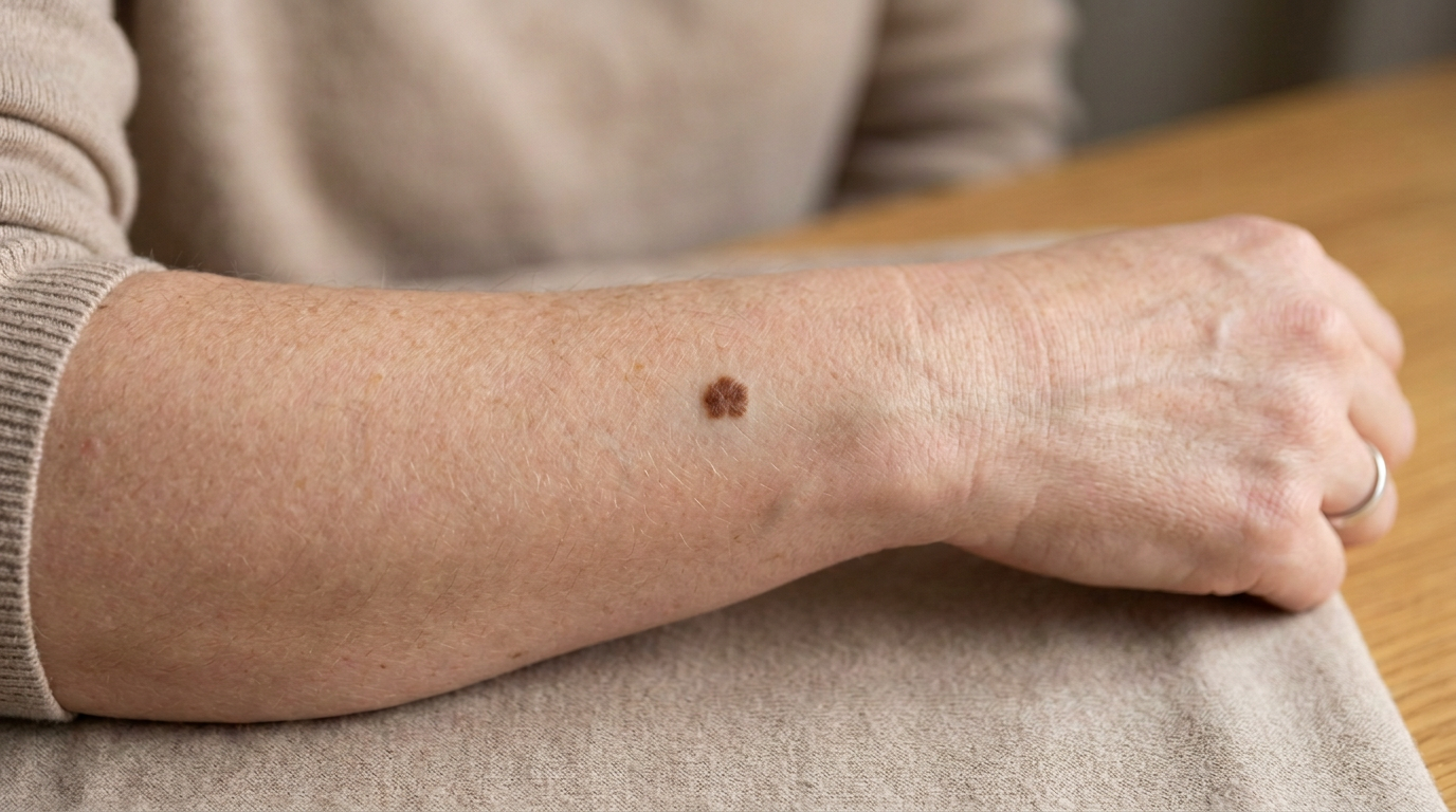
A report may show positive margins because of how the mole was removed. Many atypical moles are sampled with a shave biopsy, saucerization, or small punch biopsy rather than a wide excision. These procedures are often appropriate because they are quick, practical, and designed to answer the main diagnostic question: Is this mole benign, atypical, or melanoma? A shave biopsy may remove the visible lesion but leave microscopic nevus cells at the base or side.
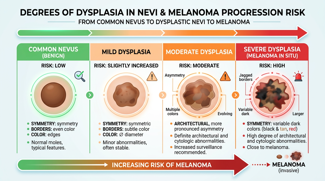
The diagnosis also depends on how the melanocytic nevus looks microscopically. Pathologists assess symmetry, nesting patterns, bridging between rete ridges, fibroplasia, inflammation, and cytologic atypia. These features help separate a common benign nevus from a dysplastic nevus and help grade the degree of dysplasia. The terms mild dysplasia, moderate dysplasia, and severe dysplasia are used to communicate how abnormal the cells and growth pattern appear.
Dysplastic nevus positive margins may also appear when the visible mole has irregular borders or extends microscopically beyond what could be seen on the skin surface. The margin status is not based on a naked-eye view alone; it is based on where cells are present on the glass slide. Sometimes the dermatologist clinically removed the entire visible mole, but the microscope still shows rare nevus cells at an edge. This is why an atypical mole biopsy report should be interpreted together with the original skin exam, dermoscopy findings, biopsy type, and any remaining pigmentation at the site.
How Serious Are Dysplastic Nevus Positive Margins?
Dysplastic nevus positive margins can be minor, meaningful, or somewhere in between, depending mainly on the degree of dysplasia. Mild dysplasia with positive margins is often managed with clinical observation if no pigment remains and the dermatologist is comfortable with the appearance of the site. Moderate dysplasia is more variable; some clinicians observe, while others recommend conservative re-excision, especially if the lesion was only partially sampled. Severe dysplasia is usually treated more cautiously because it can overlap microscopically with very early melanoma in some cases.
The key point is that dysplastic nevus positive margins is not the same as melanoma with positive margins. A dysplastic nevus is not melanoma, although severe dysplasia may prompt a pathologist to use careful language and may trigger additional treatment. Moderate dysplasia often causes the most confusion because practice patterns differ, and the decision may depend on patient risk factors, family history, lesion size, and whether pigment is still visible. Severe dysplasia at a margin often leads to a recommendation for complete excision so the entire area can be evaluated and removed.
Pathologists are especially careful when a lesion has severe dysplasia, asymmetry, pagetoid spread, or other features that raise concern for melanoma in situ. In that setting, the pathology report may include a comment recommending complete removal or clinicopathologic correlation. “Clinicopathologic correlation” means the microscope findings should be matched with the dermatologist’s view of the lesion and the patient’s history. When uncertainty remains, a second pathology opinion can be very useful; When Should You Get a Second Pathology Opinion? explains situations where another expert review may help.
What Happens Next: Treatment and Monitoring
Next steps after an atypical mole biopsy depend on the report wording and the clinical situation. If the diagnosis is mild dysplasia with positive margins, many dermatologists monitor the site during routine skin checks. If the diagnosis is moderate dysplasia, the care plan may be observation or re-excision, depending on the margin status and visible findings. If the diagnosis is severe dysplasia, many clinicians recommend excision with clear margins to reduce uncertainty and allow full evaluation.
A re-excision is different from the original biopsy. It usually removes a small rim of additional skin around the scar so any remaining atypical cells can be cleared. The tissue is then sent to pathology, where the pathologist checks whether residual dysplastic nevus is present and whether the new margins are clear. This follow-up report may say there is scar only, residual melanocytic nevus, residual dysplastic nevus, or no residual lesion.
Monitoring also matters because dysplastic nevi can be markers of increased melanoma risk, especially when numerous atypical moles or a family history of melanoma is present. A dermatologist may recommend total body skin examinations, photographs, dermoscopy, and instructions for watching changing moles. Dysplastic nevus positive margins should be considered one piece of a larger skin-risk picture, not a standalone emergency. Patients who want help reading the entire report may benefit from Understanding Your Pathology Report: How to Read It with Confidence.
Questions to Ask Your Doctor or Pathologist
Good questions can turn a confusing report into a practical plan. The goal is not to memorize pathology language, but to understand what the result means for the specific biopsy site. Patients can bring the pathology report, procedure note, and any clinical photographs to the appointment. The following questions are especially useful after dysplastic nevus positive margins appears in a report:
- What degree of dysplasia was found: mild dysplasia, moderate dysplasia, or severe dysplasia?
- Which margin is involved: peripheral, deep, or both?
- Was the original procedure a shave biopsy, punch biopsy, or excision?
- Is there any visible pigment or remaining mole at the biopsy site?
- Is re-excision recommended, or is clinical monitoring reasonable?
- Does the report show any features concerning for melanoma in situ?
- Would a dermatopathology second opinion change management in this case?
These questions help connect the pathology diagnosis to the skin exam. Positive margins on a report should not be interpreted in isolation, because the dermatologist may know whether the visible lesion was completely removed or whether residual pigment remains. Margin status has the most meaning when combined with the grade of dysplasia and the clinical impression.
A second review can be appropriate when the wording is severe, borderline, unusual, or inconsistent with the dermatologist’s concern. Expert dermatopathology review is also reasonable before a larger excision when the diagnosis could affect the size of surgery. Patients seeking practical steps can review How to Get a Second Opinion on Your Pathology Diagnosis.
Frequently Asked Questions
Does dysplastic nevus positive margins mean cancer?
No. Dysplastic nevus positive margins means atypical mole cells reach the edge of the biopsy sample, not that cancer has been diagnosed. A dysplastic nevus is different from melanoma, although severe dysplasia can sometimes require careful distinction from early melanoma. The report should be reviewed for the exact degree of dysplasia and any comment about melanoma in situ.
Do positive margins always need another excision?
No. Positive margins do not always require another excision. Mild dysplasia is often observed if the biopsy site looks clear clinically. Moderate dysplasia may be observed or re-excised depending on the dermatologist’s assessment. Severe dysplasia is more often treated with complete excision.
What is the difference between moderate dysplasia and severe dysplasia?
Moderate dysplasia means the mole cells and growth pattern are more atypical than mild dysplasia but not at the highest level of concern. Severe dysplasia means the changes are more pronounced and may overlap with melanoma in situ in some features. Because of that overlap, severe dysplasia is usually managed more aggressively. The distinction can sometimes benefit from dermatopathology review.
Can dysplastic nevus positive margins come back later?
Yes, dysplastic nevus positive margins can be followed by recurrent pigmentation at the scar if nevus cells remain. This is often called a recurrent nevus phenomenon and can look concerning on the skin. Any new or changing pigment in a biopsy scar should be examined by a dermatologist. Sometimes another biopsy is needed to separate recurrence from melanoma.
Should I get a second opinion on an atypical mole biopsy?
A second opinion can be helpful when an atypical mole biopsy shows severe dysplasia, borderline features, or wording that is difficult to understand. It can also help when the recommended treatment feels more extensive than expected. A second opinion does not mean the first diagnosis was wrong; it means another qualified pathologist reviews the slides. This can provide reassurance or refine the diagnosis before treatment.
A report saying dysplastic nevus positive margins can feel unsettling, but it is a common and manageable situation. The most useful next step is to connect the margin finding with the dysplasia grade, the biopsy type, and the dermatologist’s exam. Honest Pathology consultations can help patients and families understand the wording of a report and prepare focused questions for the treating clinician.
References:
National Cancer Institute — Pathology Reports
National Cancer Institute — Dysplastic Nevus
MedlinePlus — Moles