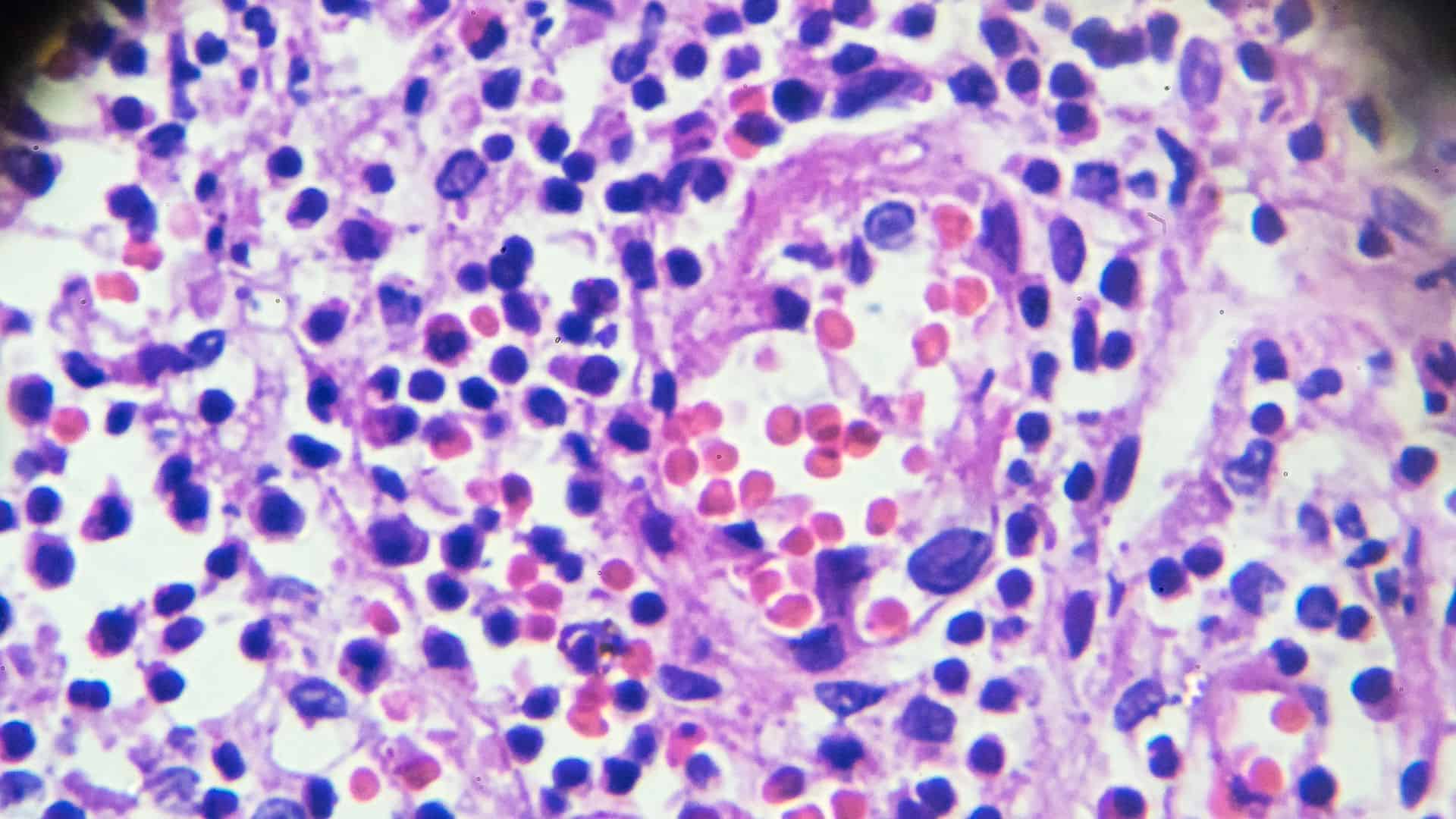
Understanding Your Breast Papilloma Diagnosis
Clear Guidance for Interpreting Your Pathology Results
A breast papilloma is a noncancerous growth that develops within the milk ducts of the breast. These growths are often discovered during imaging studies, evaluation of nipple discharge, or biopsy procedures. Your pathology report helps determine the type of papilloma present and whether any additional tissue changes require further assessment.
HONEST Pathology specialists carefully evaluate breast tissue samples and provide clear, patient-friendly explanations of your findings. We help you better understand your diagnosis, possible next steps, and any recommendations from your healthcare team so you can make informed decisions about your breast health.

Informed Decisions

Better Conversations

Leave Empowered
Ready to Understand Your Diagnosis?
Don’t let confusion hold you back. Schedule an online consultation with our expert pathologists and get clear, personalized answers about your diagnosis — all from the comfort of home.
Frequently Asked Questions
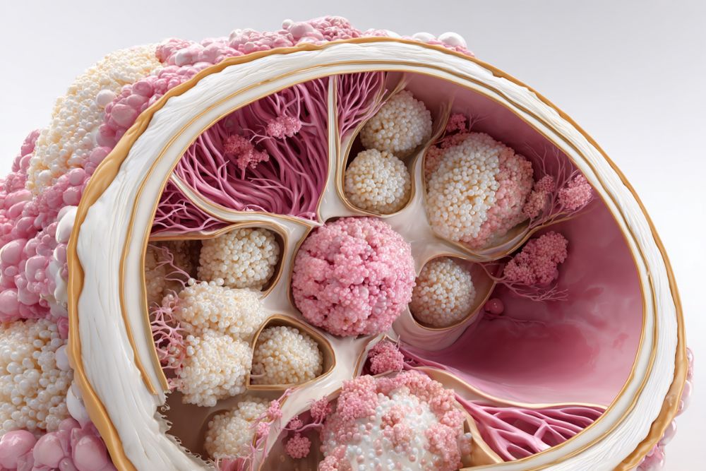
A breast papilloma is a benign tumor that forms inside a breast duct. It consists of glandular and fibrous tissue along with blood vessels.
No. Most breast papillomas are noncancerous growths. However, some cases may contain atypical cells that require additional evaluation.
Some patients experience nipple discharge, a small lump near the nipple, or breast discomfort, while others have no noticeable symptoms.
Breast papillomas are commonly identified through mammograms, ultrasound imaging, breast MRI, or tissue biopsy.
A solitary papilloma refers to a single papilloma, usually found in one of the larger ducts near the nipple area.
Yes. Some individuals develop multiple papillomas in smaller ducts, which may require closer monitoring depending on the findings.
A biopsy allows the pathologist to examine the tissue under a microscope and confirm whether the growth is benign or associated with other cellular changes.
Your healthcare provider may recommend observation, surgical removal, or additional imaging based on the pathology findings and your individual clinical situation.