Understanding Your Bladder Cancer Diagnosis
Clear, Supportive Insights for Your Diagnostic Findings
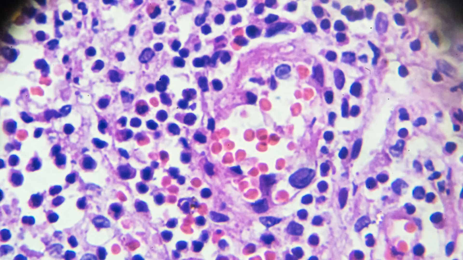
Bladder cancer begins in the lining of the bladder and varies in cell type, grade, and depth of invasion. Your pathology report outlines these features clearly, helping you and your care team make informed decisions about treatment and ongoing management.
HONEST Pathology specialists walk you through each part of your bladder cancer report, offering clear explanations and supportive guidance. We help you understand what every detail means so you feel confident in your diagnosis and next steps.


Informed Decisions

Better Conversations

Leave Empowered

Peace of Mind

Guided Next Steps
Ready to Understand Your Diagnosis?
Don’t let confusion hold you back. Schedule an online consultation with our expert pathologists and get clear, personalized answers about your diagnosis — all from the comfort of home.
Frequently Asked Questions
Bladder cancers can include urothelial carcinoma, squamous cell carcinoma, or adenocarcinoma. Each type responds differently to treatments, so identifying the tumor type helps your care team select the most appropriate therapy.
Tumor grade describes how abnormal the cancer cells look under the microscope. High-grade tumors grow more aggressively and may require more intensive treatment, while low-grade tumors tend to behave more slowly.
Pathologists assess how deeply the tumor extends into the bladder wall. Tumors limited to the surface layers may be treated with local therapies, while those invading deeper muscle layers often require more extensive treatment.
Carcinoma in situ (CIS) is a flat, high-grade cancer that can spread quickly. Its detection may change treatment plans and increase the need for close monitoring or additional therapies.
Yes. Testing for genetic changes such as FGFR3 mutations can help identify targeted treatments that may improve outcomes for some patients.
Lymphovascular invasion indicates that cancer cells have entered the lymphatic or blood vessels. This finding may increase the likelihood of cancer spread and influence treatment recommendations.
Clear margins mean no cancer cells were found at the edges of the removed tissue. Positive margins may indicate residual disease and can guide decisions about additional treatment.
Lymph nodes removed during surgery are examined under the microscope. Identifying cancer cells in lymph nodes helps stage the disease and plays a major role in planning treatment strategies.