

A frightening pancreatic report can feel unclear. pancreatic FNA atypical cells may not be final cancer; learn what to ask next.
Pancreatic FNA atypical cells means the sample contains cells that look abnormal, but the pathologist may not have enough certainty to make a definite cancer diagnosis. This wording often appears on a cytology report after a fine needle aspiration of a pancreatic mass, cyst, or duct-related abnormality. It can also appear with similar language, such as suspicious cells, atypical ductal cells, or suspicious for malignancy.
Receiving this result can feel especially frightening because the pancreas is a difficult organ to sample and pancreatic cancer is a diagnosis many patients fear. The word “atypical” can sound like a warning without giving a clear answer. In reality, pancreatic FNA atypical cells is a gray-zone result, not a complete explanation by itself. The next step is usually correlation with imaging, symptoms, blood tests, and sometimes repeat sampling or expert review.
Pancreatic FNA Atypical Cells — What It Actually Means
Pancreatic FNA atypical cells are abnormal-looking cells seen in a sample collected from the pancreas using a thin needle. The sample is usually obtained during endoscopic ultrasound, when a gastroenterologist guides a needle into a pancreatic mass or suspicious area. The pathologist then examines the cells under a microscope and decides whether they are benign, atypical, suspicious, malignant, or nondiagnostic. A pancreatic biopsy result in this setting is often based on tiny clusters of cells rather than a large piece of tissue.
A helpful analogy is looking at a few puzzle pieces without seeing the whole picture. Some pieces may clearly show a blue sky, a face, or a building; others may be damaged, folded, or too few to interpret with confidence. In the pancreas, inflammation, scarring, chronic pancreatitis, treatment effect, and sampling limits can make cells look abnormal. That is why a cytology report may use cautious wording instead of giving a definite diagnosis.
The term atypical does not mean normal, but it also does not automatically mean cancer. Suspicious cells means the pathologist sees features that raise stronger concern, but the available material may still fall short of the threshold for a definitive malignant diagnosis. Fine needle aspiration samples are interpreted in context, including imaging findings, the size and location of the pancreatic mass, and whether a cell block was available for additional testing. Patients who want the basics of report structure may benefit from reviewing what a pathology report is before the follow-up appointment.
Why Your Report Shows This Finding
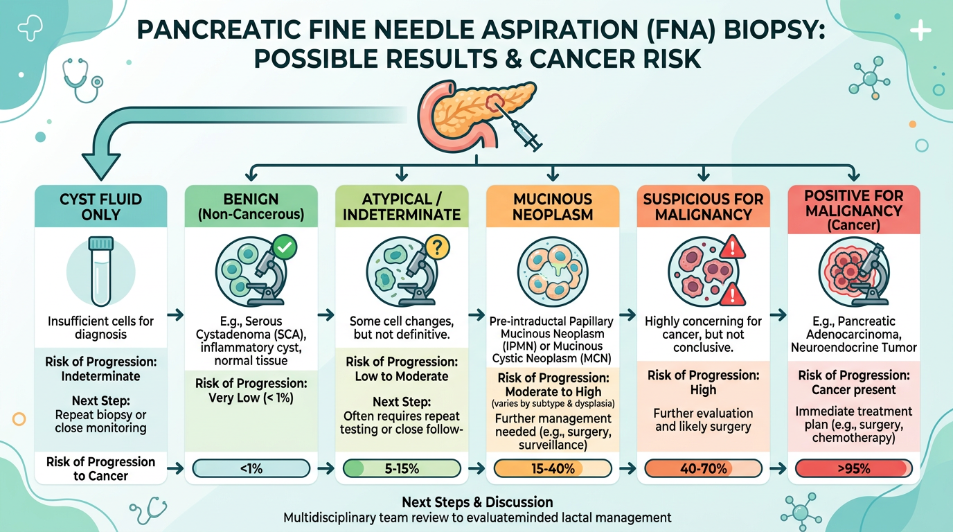
A pancreatic FNA may show atypical cells for several reasons. One common reason is that the needle collected only a small number of abnormal cells, making the pancreatic biopsy result limited. Another reason is that inflammation can distort pancreatic duct cells, especially in chronic pancreatitis or after a blocked duct. A cytology report tries to separate reactive change from ductal adenocarcinoma, but that separation can be difficult when the sample is scant.
Pathologists look at nuclear size, nuclear shape, chromatin pattern, crowding, necrosis, and the way cells form clusters. In ductal adenocarcinoma, cells often show irregular nuclei, abnormal architecture, and loss of the orderly honeycomb pattern seen in benign duct cells. Suspicious cells may show several of these features, but not enough for a firm diagnosis. A cell block can be valuable because it preserves collected material in a paraffin block, allowing special stains, immunohistochemistry, or molecular testing in selected cases.
Sometimes the report reflects a mismatch between imaging and the microscope findings. Imaging may show a pancreatic mass that looks worrisome, but the fine needle aspiration sample may contain mostly blood, gastrointestinal contamination, fibrosis, or inflammation. In other cases, the sample may capture the edge of a tumor rather than its most diagnostic center. The phrase pancreatic FNA atypical cells tells the clinical team that the sample deserves attention, correlation, and often a plan for clarification.
How Serious Are Pancreatic FNA Atypical Cells?
Pancreatic FNA atypical cells should be taken seriously, but the level of concern depends on the entire clinical picture. A small number of mildly atypical cells in a patient with pancreatitis is different from markedly suspicious cells from a solid pancreatic mass with a blocked bile duct. The seriousness also changes when imaging shows vascular involvement, weight loss, jaundice, or a rising CA 19-9 level. A pancreatic biopsy result is one piece of evidence, not the whole case.
In many institutions, cytology categories carry different estimated risks of malignancy. “Atypical” usually means an intermediate-risk category, while “suspicious for malignancy” carries a higher risk and may be managed much like cancer if imaging strongly supports that diagnosis. However, the exact risk varies by institution, sampling method, pathologist experience, and whether the finding is from a cyst, duct brushing, or solid mass. When suspicious cells are mentioned, the treating team usually moves quickly to decide whether repeat biopsy, surgical consultation, oncology evaluation, or surveillance is most appropriate.
The most important point is that pancreatic FNA atypical cells are not a final staging diagnosis. A report cannot determine treatment by wording alone unless it clearly establishes malignancy and the clinical team has enough information to plan safely. A second review can be helpful when the cytology report is ambiguous, the treatment recommendation is major, or the microscopic findings and scans do not seem to match. Patients considering this step may find practical context in when to get a second pathology opinion.
What Happens Next: Treatment and Monitoring
After a cytology report shows atypical or suspicious cells, the next step is usually a multidisciplinary discussion. Gastroenterology, radiology, surgery, oncology, and pathology may review the pancreatic mass together. The team compares the microscopic findings with CT, MRI, endoscopic ultrasound, blood tests, symptoms, and procedure notes. This comparison often determines whether the next best step is repeat fine needle aspiration, core biopsy, surgery, close imaging follow-up, or oncology referral.
A repeat procedure may be recommended if the pancreatic biopsy result is not definitive and treatment decisions depend on certainty. During repeat sampling, the proceduralist may target a different area, use a core needle, obtain more passes, or request rapid on-site evaluation if available. A better cell block may allow immunostains or molecular tests that were not possible the first time. If suspicious cells are abundant and the imaging is classic for ductal adenocarcinoma, the team may decide that additional tissue is not necessary before moving forward, depending on the planned treatment.
Monitoring can include repeat imaging, liver tests, bilirubin levels, pancreatic enzymes, nutritional assessment, and sometimes CA 19-9. For cystic lesions, fluid markers, cyst features, and interval growth may matter more than a single atypical cell cluster. For solid lesions, persistent concern usually leads to more urgent clarification. The goal is not to ignore uncertainty, but to resolve it with the safest and most informative next step.
Questions to Ask Your Doctor or Pathologist
- Does the cytology report say “atypical,” “suspicious for malignancy,” or “positive for malignancy”?
- Was the sample from a solid pancreatic mass, a cyst, a duct brushing, or a lymph node?
- Was there enough material for a cell block, immunohistochemistry, or molecular testing?
- Do the imaging findings match the pancreatic biopsy result?
- Could pancreatitis, scarring, or a prior procedure explain the atypical cells?
- Would repeat fine needle aspiration or a core biopsy likely change management?
- Should the slides be reviewed by a cytopathologist or pancreatic pathology specialist?
These questions help turn vague wording into a practical plan. A report that says pancreatic FNA atypical cells can be unsettling, but many reports contain clues about sample adequacy, diagnostic confidence, and recommended correlation. Reading the diagnosis, comment, adequacy statement, and procedure details together can make the result less confusing. A step-by-step approach to report language is available in understanding a pathology report.
A pathology consultation can be especially useful when treatment choices are high stakes. This review typically confirms the diagnosis, explains the uncertainty, and identifies whether additional stains, levels, or repeat sampling could help. For patients preparing records, how to get a second opinion on a pathology diagnosis outlines the practical steps.
Frequently Asked Questions
Does atypical cells on pancreatic FNA mean cancer?
No, atypical cells on pancreatic FNA does not automatically mean cancer. Pancreatic FNA atypical cells means the cells look abnormal, but the pathologist may not have enough evidence for a definitive malignant diagnosis. The risk is higher when imaging shows a solid pancreatic mass or when the report says suspicious cells. The clinical team usually combines the cytology report with imaging and symptoms before deciding what comes next.
What does suspicious for malignancy mean on a pancreatic biopsy?
Suspicious for malignancy means the pathologist sees features strongly concerning for cancer, but the sample may still be limited. This wording is more concerning than atypical, especially when the pancreatic biopsy result comes from a solid mass. Suspicious cells often lead to repeat sampling, specialist review, or treatment planning based on the whole case. The exact next step depends on imaging, surgical options, and whether more tissue is needed.
Can a pancreatic FNA be wrong?
Yes, a pancreatic FNA can be limited or occasionally misleading because the pancreas is difficult to sample. A fine needle aspiration may miss the most diagnostic part of a tumor or collect too few cells. Inflammation and chronic pancreatitis can also mimic some cancer-like changes. That is why a cytology report with pancreatic FNA atypical cells often triggers correlation or repeat biopsy rather than an immediate final conclusion.
Why would my doctor repeat the pancreatic biopsy?
A repeat biopsy may be recommended when the first sample did not provide enough certainty for treatment decisions. The goal is to obtain more diagnostic cells, a better cell block, or tissue for additional testing. A repeat procedure may target a different area of the pancreatic mass or use a core biopsy needle. This can help separate inflammation from ductal adenocarcinoma when the first cytology report is unclear.
Should I get a second opinion for atypical pancreatic cells?
A second opinion is reasonable when a report is ambiguous, when major surgery or chemotherapy is being considered, or when imaging and pathology do not seem to match. Pancreatic FNA atypical cells can be reviewed by a cytopathologist with pancreatic experience. The review may confirm the original interpretation, refine the category, or recommend more testing. It can also give the treating team clearer language for planning the next step.
Ambiguous pancreatic cytology can feel like being left between answers, but uncertainty in a report is often a signal to gather better evidence, not a reason to assume the worst. Honest Pathology consultations can help clarify what the wording means, whether the sample was adequate, and what questions may be most useful before treatment decisions are made.
References:
National Cancer Institute — Pathology Reports
National Cancer Institute — Fine-Needle Aspiration Biopsy
National Cancer Institute — Pancreatic Cancer Treatment
