

Fear after a colonoscopy report is normal. Intramucosal carcinoma colon polyp findings have specific meaning. Learn what to ask next.
Intramucosal carcinoma colon polyp usually means cancer-like cells were found only within the inner lining of a colon polyp, without clear invasion into the deeper tissue where spread becomes possible. This diagnosis can sound frightening because the word carcinoma appears in the pathology report. In the colon, however, location matters as much as the name of the abnormal cells.
Many patients see this phrase after a colonoscopy and immediately worry about invasive colon cancer, chemotherapy, or surgery. That fear is understandable. Pathologists use precise language because small differences under the microscope can change treatment decisions, but the wording can feel cold and alarming when read without an explanation.
The most reassuring point is that an intramucosal carcinoma colon polyp is often managed very differently from a deeply invasive cancer. The key questions are whether the polyp removal was complete, whether the abnormal cells reached the stalk or margin, and whether there is any true invasion beyond the mucosa.
Intramucosal Carcinoma Colon Polyp — What It Actually Means
An intramucosal carcinoma colon polyp is a polyp in which the most abnormal cells are confined to the mucosa, the thin inner lining of the colon. The mucosa is like the wallpaper inside a room: It can develop serious surface damage, but that damage has not necessarily broken through into the deeper wall. In this setting, the abnormal cells may look very concerning, yet they remain in a compartment that lacks the same access to lymphatic channels found deeper in the colon wall.
During a colon polyp biopsy or polyp removal, the tissue is processed into very thin sections and examined under a microscope. Pathologists look at the shape of the glands, the appearance of the cell nuclei, and how far the abnormal growth extends. A pathology report may use terms such as high-grade dysplasia, carcinoma in situ, or intramucosal carcinoma when the lesion is still limited to the mucosal layer. These terms overlap in daily practice, but they all point to a noninvasive or preinvasive category in the colon when no deeper invasion is identified.
The distinction matters because invasive colon cancer requires tumor cells to grow beyond the muscularis mucosae into the submucosa. The muscularis mucosae is a very thin muscle layer beneath the mucosa, and crossing it changes the risk profile. If the abnormal cells remain above that layer, lymph node metastasis is considered extremely unlikely because the mucosa has very limited lymphatic drainage. This is why an intramucosal carcinoma colon polyp can sound like cancer but behave more like an advanced polyp when completely removed.
Why Your Report Shows This Finding
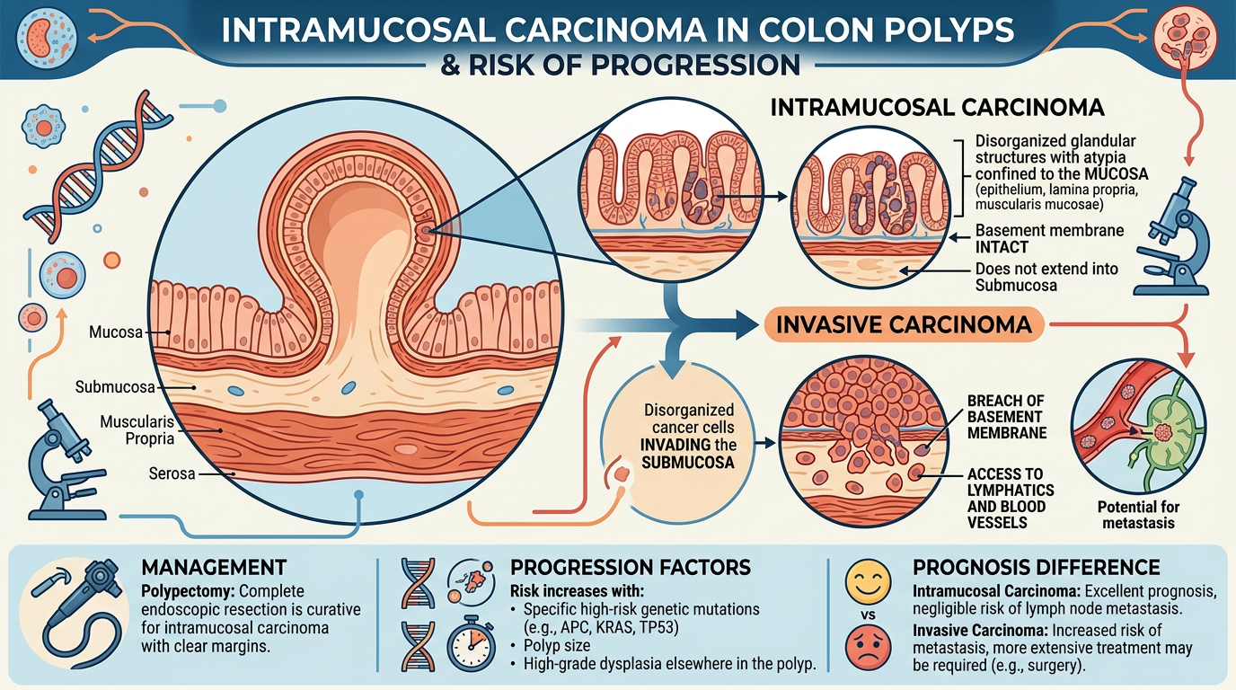
This finding usually develops within an adenoma, which is a common type of precancerous colon polyp. Over time, some adenomas can accumulate genetic changes that make the cells more disorganized and aggressive-looking. The sequence may progress from low-grade dysplasia to high-grade dysplasia and, in some reports, intramucosal carcinoma. A colon polyp biopsy captures a sample of this process, while complete polyp removal allows the pathologist to see the full architecture and deepest extent.
Under the microscope, pathologists search carefully for invasion beyond the mucosa. The lamina propria is the delicate supportive tissue between the glands in the mucosal layer. Tumor cells within the lamina propria may justify the term intramucosal carcinoma, but that is still different from invasion into the submucosa. The muscularis mucosae is the boundary that helps separate these categories, although it can be distorted or hard to see in large, cauterized, or fragmented polyps.
Several technical details can affect how confident the diagnosis appears in the pathology report. A polyp removed in one piece is usually easier to evaluate than a polyp removed in fragments. Cautery artifact, which is heat-related distortion from removal, can blur the edge of the lesion. If the pathologist cannot clearly assess the margin, the report may recommend repeat colonoscopy, close endoscopic follow-up, or additional clinical correlation to confirm that the polyp removal was complete.
How Serious Is an Intramucosal Carcinoma Colon Polyp?
An intramucosal carcinoma colon polyp is serious enough to deserve careful attention, but it is not automatically the same as invasive colon cancer. The most important issue is whether the abnormal cells were completely removed and whether there is any evidence that they crossed into the submucosa. When the diagnosis is truly limited to the mucosa and the margins are clear, many patients do not need colon surgery. Instead, care often focuses on complete endoscopic removal and surveillance colonoscopy.
The reason the outlook is usually favorable is tied to anatomy. Lymph node metastasis from a purely mucosal colon lesion is considered essentially absent or extraordinarily rare because the relevant lymphatic channels are found mainly below the muscularis mucosae. This is why pathologists spend so much time evaluating the depth of invasion. In contrast, invasive colon cancer begins when malignant glands enter the submucosa, where access to lymphatic and blood vessels becomes more clinically meaningful.
The level of concern rises when the specimen is fragmented, the cauterized margin is involved, or the report cannot exclude deeper invasion. In those situations, an intramucosal carcinoma colon polyp may still be curable, but the treating gastroenterologist may recommend another procedure to make sure no residual lesion remains. If a report mentions submucosal invasion, lymphovascular invasion, poor differentiation, tumor budding, or an involved deep margin, the discussion changes because those features can be associated with invasive colon cancer. For patients trying to understand risk, the phrase itself is only the starting point; the margin status, depth, and specimen quality provide the real clinical context.
What Happens Next: Treatment And Monitoring
Next steps usually begin with the gastroenterologist reviewing the colonoscopy findings and the pathology report together. A small polyp that was removed completely in one piece may need only surveillance at an interval based on national guidelines and the patient’s full polyp history. A larger polyp, a piecemeal polyp removal, or an uncertain margin may lead to a repeat colonoscopy sooner. The goal is to confirm that no residual abnormal tissue is left behind.
Some patients are referred to a colorectal surgeon, but referral does not always mean surgery is required. It often means the team wants expert input about whether endoscopic treatment was enough. If the lesion is truly an intramucosal carcinoma colon polyp with no submucosal invasion, no lymphovascular invasion, and no high-risk margin issue, colectomy is commonly avoided. If invasive colon cancer is identified, then staging, imaging, and lymph node evaluation may become part of care.
Patients may also benefit from requesting the full report, not just the summary line. Resources such as What Is a Pathology Report? and Understanding Your Pathology Report: How to Read It with Confidence can make the terminology easier to follow. A pathology report is not only a diagnosis; it is a map of what was seen, how complete the polyp removal appears, and what uncertainties remain. That map helps the care team choose between routine surveillance, early repeat colonoscopy, expert endoscopic removal, or surgical consultation.
Questions To Ask Your Doctor Or Pathologist
- Was the intramucosal carcinoma colon polyp removed completely in one piece or in fragments?
- Does the pathology report show clear margins, involved margins, or margins that cannot be assessed?
- Did the pathologist see any submucosal invasion, or is the lesion confined to the mucosa?
- Is the diagnosis closer to high-grade dysplasia, carcinoma in situ, or intramucosal carcinoma?
- Was lymphovascular invasion mentioned, or was it absent?
- When should the next colonoscopy be performed after this polyp removal?
- Would a second pathology review change management in this specific case?
These questions help turn a frightening phrase into practical next steps. A colon polyp biopsy or excision report often contains enough information to estimate risk, but the wording may require translation into plain language. Patients should feel comfortable asking whether the diagnosis means noninvasive disease, invasive colon cancer, or an uncertain borderline finding.
A second review can be helpful when the polyp was large, fragmented, cauterized, or described with uncertain invasion. Guidance on When Should You Get a Second Pathology Opinion? and How to Get a Second Opinion on Your Pathology Diagnosis may help patients prepare records and slides for review. A second opinion is especially useful when treatment could shift from surveillance to surgery.
Frequently Asked Questions
Is intramucosal carcinoma in a colon polyp cancer?
Intramucosal carcinoma colon polyp is a diagnosis with cancer-like cells limited to the inner mucosal layer of the colon. In the colon, many clinicians treat this as a noninvasive or very early lesion when no submucosal invasion is present. The word carcinoma is alarming, but the depth of invasion is the deciding factor. The report should be reviewed for margins and any mention of invasive colon cancer.
Can intramucosal carcinoma in a colon polyp spread?
A true intramucosal carcinoma colon polyp is not expected to spread to lymph nodes because the abnormal cells have not reached the submucosa. Lymph node metastasis becomes a concern when cancer invades deeper tissue where lymphatic channels are present. If the report mentions only lamina propria involvement and no submucosal invasion, the risk is usually very low. Unclear margins or fragmented tissue may still require follow-up.
Do I need surgery for intramucosal carcinoma in a colon polyp?
Surgery is not always needed for this diagnosis. If polyp removal was complete and the abnormal cells are limited to the mucosa, surveillance colonoscopy may be the main plan. Surgery may be discussed if invasive colon cancer is found, if the margin is involved, or if the lesion cannot be fully removed endoscopically. The decision depends on the entire pathology report and colonoscopy findings.
What does margin mean on my colon polyp pathology report?
The margin is the cut or cauterized edge of the removed tissue. A clear margin means the abnormal area appears fully removed in the examined sections. An involved or uncertain margin means abnormal cells are at the edge or the edge cannot be reliably assessed. In that setting, repeat colonoscopy or additional polyp removal may be recommended.
Should I get a second opinion on intramucosal carcinoma?
A second opinion can be reasonable when the diagnosis affects whether a patient needs surgery or surveillance. It is especially useful if the colon polyp biopsy was fragmented, the muscularis mucosae is hard to identify, or the report says invasion cannot be excluded. Another gastrointestinal pathologist may confirm the diagnosis or clarify the depth of invasion. This review can help the treating team choose the safest next step.
An intramucosal carcinoma colon polyp can feel overwhelming, but the most meaningful details are usually visible in the report: depth, margins, and completeness of removal. Many cases are managed successfully with complete endoscopic treatment and careful follow-up. Honest Pathology consultations can help patients and families understand the wording, organize questions, and decide whether expert slide review is appropriate.
References:
National Cancer Institute — Pathology Reports
National Cancer Institute — Carcinoma In Situ
National Cancer Institute — Adenoma
