

A scary biopsy result can feel overwhelming. atypical endometrial hyperplasia has clear next steps; learn what to ask next.
atypical endometrial hyperplasia means that cells in the uterine lining have become crowded, irregular, and concerning enough that doctors treat the finding seriously. This diagnosis is often closely related to the term EIN, which stands for endometrial intraepithelial neoplasia. Both terms describe a precancerous change in the endometrium, the tissue that normally thickens and sheds with menstrual cycles.
Seeing words like “atypical,” “neoplasia,” or “precancer” on an endometrial biopsy report can feel frightening. Many patients immediately wonder whether cancer is already present, whether surgery is unavoidable, and whether time has been lost. The most helpful first step is to understand what the pathologist actually saw under the microscope and why the diagnosis leads to careful follow-up.
This result does not mean that every patient already has cancer. It does mean that the uterine lining has changed in a way that deserves prompt, organized medical attention.
Atypical Endometrial Hyperplasia — What It Actually Means
Atypical endometrial hyperplasia is a diagnosis made by examining tissue from the uterine lining under a microscope. The word “hyperplasia” means that there are more glands than expected, and the word “atypical” means that the cells look abnormal in size, shape, or organization. In many modern reports, the same process may be called an EIN diagnosis, because EIN better describes a clonal, precancerous growth pattern. The two terms are often used together because they point to the same practical concern: the tissue has a higher risk of being associated with, or later developing into, endometrial cancer.
A useful analogy is a garden where some plants have started growing too densely and in an unusual pattern. The garden is not necessarily overrun, but the pattern tells the gardener that a closer look is needed. In the same way, an endometrial biopsy samples part of the uterine lining and allows the pathologist to study the architecture of the glands and the appearance of the cells. If the glands are crowded and the cells look atypical, atypical endometrial hyperplasia becomes a serious diagnostic consideration.
The report may also mention “complex atypical hyperplasia,” “EIN,” or “cannot exclude well-differentiated endometrioid adenocarcinoma.” These phrases can sound very different, but they often sit along the same diagnostic spectrum. A pathology report is built from microscopic patterns, clinical information, and sometimes additional studies. Pathologists often review the endometrial biopsy carefully because separating atypical endometrial hyperplasia from early endometrial cancer can be challenging, especially when the sample is small or fragmented.
Why The Report Shows This Finding
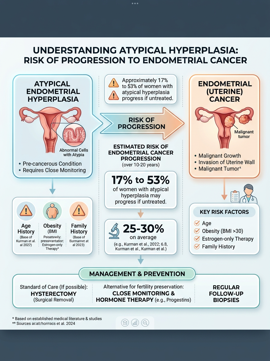
Atypical endometrial hyperplasia usually develops when the uterine lining is exposed to estrogen stimulation that is not adequately balanced by progesterone. This hormonal environment can occur with irregular ovulation, polycystic ovary syndrome, obesity, perimenopause, estrogen therapy without progesterone, or certain ovarian conditions. Abnormal bleeding is one of the most common reasons an endometrial biopsy is performed, especially bleeding after menopause or heavy, irregular bleeding before menopause. The biopsy is intended to answer whether the uterine lining is benign, overgrown, precancerous, or malignant.
Under the microscope, the pathologist looks at both the architecture and the cytology. Architecture refers to how crowded and complex the glands are, while cytology refers to how abnormal the individual cells look. In an EIN diagnosis, the glands are usually crowded compared with the surrounding background endometrium, and the cells look different from nearby benign glands. The diagnosis is not made from one isolated cell; it is made from a pattern across the tissue.
Sometimes the report shows atypical endometrial hyperplasia because the biopsy caught only part of a larger process. An endometrial biopsy is a sample, not a complete map of the uterus. That is why a hysterectomy specimen may later show more tissue changes than were visible in the original biopsy. The presence of abnormal bleeding, risk factors, and biopsy findings together helps the gynecologist decide whether further sampling, progesterone therapy, or hysterectomy should be discussed.
How Serious Is Atypical Endometrial Hyperplasia?
Atypical endometrial hyperplasia is serious because it carries a meaningful risk of endometrial cancer, either already present elsewhere in the uterus or developing later if untreated. Published studies have shown that a substantial minority of patients diagnosed with EIN or atypical hyperplasia on biopsy have cancer found when the uterus is later removed. This does not mean that cancer is guaranteed. It means the diagnosis is significant enough that careful treatment planning is warranted.
The seriousness depends on several details, including age, menopausal status, bleeding pattern, medical conditions, fertility goals, imaging findings, and the exact wording of the pathology report. For a postmenopausal patient with abnormal bleeding, atypical endometrial hyperplasia often raises stronger concern than the same phrase might in a younger patient with known hormonal imbalance. However, the diagnosis should never be ignored in either situation. Endometrial cancer can sometimes be well differentiated and subtle, making expert review of the endometrial biopsy valuable when the wording is borderline.
In practical terms, atypical endometrial hyperplasia sits in a “high attention” category, not a “panic” category. The finding gives clinicians a chance to act before an invasive cancer develops or while a very early cancer may still be highly treatable. Some patients are treated with hysterectomy, while carefully selected patients may receive progesterone therapy with close monitoring. When the report language is uncertain, a second pathology opinion can help confirm whether the biopsy truly shows atypical endometrial hyperplasia, benign hyperplasia without atypia, or possible endometrial cancer.
What Happens Next: Treatment And Monitoring
After an endometrial biopsy shows atypical endometrial hyperplasia, the next step is usually a detailed discussion with a gynecologist or gynecologic oncologist. Many patients are referred to a gynecologic oncologist because this specialist routinely manages precancerous and cancerous conditions of the uterine lining. The most definitive treatment is hysterectomy, which removes the uterus and allows the entire lining to be examined. The hysterectomy specimen can confirm the biopsy diagnosis and determine whether endometrial cancer is also present.
For patients who are not ready for surgery, are poor surgical candidates, or strongly desire future pregnancy, progesterone therapy may be considered. Progesterone therapy can be given as an intrauterine device, oral medication, or another regimen chosen by the treating clinician. This approach requires close follow-up because the abnormal tissue must be monitored to make sure it regresses rather than persists or progresses. Repeat endometrial biopsy is commonly used to assess response after several months of treatment.
Monitoring is not a passive approach. It is an active plan with timelines, repeat tissue sampling, and clear decision points. If atypical endometrial hyperplasia persists despite progesterone therapy, or if endometrial cancer is found, the treatment plan may change. Patients often benefit from bringing the full pathology report, imaging results, medication list, and bleeding history to the consultation. A plain-language resource on understanding a pathology report can also help patients prepare for that visit with more confidence.
Questions To Ask Your Doctor Or Pathologist
- Does the report use the term EIN diagnosis, atypical endometrial hyperplasia, complex atypical hyperplasia, or possible carcinoma?
- Was the endometrial biopsy sample adequate, limited, fragmented, or difficult to interpret?
- Is there any wording that suggests possible endometrial cancer already present in the sample?
- Should the pathology slides be reviewed by a gynecologic pathologist before treatment decisions are finalized?
- Is hysterectomy recommended, or is progesterone therapy a reasonable option in this situation?
- How soon should repeat sampling be done if nonsurgical treatment is chosen?
- What symptoms, especially abnormal bleeding, should prompt earlier follow-up?
These questions help turn a frightening phrase into a practical conversation. Atypical endometrial hyperplasia is not just a laboratory label; it is a finding that connects the microscopic appearance of the uterine lining with real decisions about treatment and monitoring.
When the diagnosis will affect fertility, surgery, or cancer treatment planning, a second review can be especially helpful. Patients can ask whether the original slides can be sent for another opinion, and a practical overview of how to get a second opinion may make that process feel less intimidating.
Frequently Asked Questions
Is atypical endometrial hyperplasia cancer?
Atypical endometrial hyperplasia is considered a precancerous diagnosis, not automatically cancer. However, it is closely associated with endometrial cancer, and cancer may sometimes be found elsewhere in the uterus when more tissue is examined. That is why doctors take the diagnosis seriously and often recommend definitive treatment or close monitoring. The endometrial biopsy result should be discussed promptly with the treating gynecologist.
What does EIN mean on an endometrial biopsy?
EIN means endometrial intraepithelial neoplasia, a term used for a precancerous glandular growth in the uterine lining. An EIN diagnosis often overlaps with atypical endometrial hyperplasia in clinical practice. The term helps identify lesions that have a higher risk of progression or associated carcinoma. The exact wording of the report may guide whether treatment, repeat sampling, or expert pathology review is recommended.
Can atypical endometrial hyperplasia go away with progesterone?
Atypical endometrial hyperplasia can regress with progesterone therapy in selected patients. This option is most often considered when surgery is risky or when fertility preservation is a major goal. Close follow-up is essential because the uterine lining must be resampled to confirm response. If the abnormality persists or progresses, treatment recommendations usually change.
Why is hysterectomy recommended for EIN?
Hysterectomy is often recommended because EIN and atypical endometrial hyperplasia can coexist with endometrial cancer that was not captured in the biopsy sample. Removing the uterus treats the precancerous process and allows the entire hysterectomy specimen to be examined. This complete evaluation can reveal whether cancer is present and whether additional treatment is needed. The recommendation may differ for patients who need fertility-sparing care or cannot safely undergo surgery.
Should I get a second opinion for atypical endometrial hyperplasia?
A second opinion can be reasonable when atypical endometrial hyperplasia is unexpected, borderline, or likely to change major treatment decisions. Gynecologic pathology can be nuanced because benign hyperplasia, EIN diagnosis, and early endometrial cancer may look similar in small samples. A second review typically examines the original slides rather than requiring a new biopsy. The goal is diagnostic confidence before decisions such as hysterectomy or progesterone therapy are made.
A diagnosis involving the uterine lining can feel deeply personal and unsettling, but clarity is possible. The key is to understand the exact biopsy wording, the risk being addressed, and the next medical step. Honest Pathology consultations can help patients and caregivers understand whether the pathology language is clear, whether a second opinion may be useful, and what questions should be brought to the treating team.
References:
National Cancer Institute — Pathology Reports Fact Sheet
National Cancer Institute — Endometrial Hyperplasia Definition
MedlinePlus — Endometrial Cancer
