1
Colonoscopy
A clinician examines the inside of the colon and identifies tissue that may require sampling.

Get a clearer explanation of the diagnosis, tumour type, depth of invasion, lymph nodes, margins, high-risk features, and biomarker results documented in your colon pathology report.
Not sure which service you need? Compare your options →This consultation helps you understand existing pathology results and prepare questions for your care team. It does not replace diagnosis, complete staging, or treatment planning by your treating clinicians.
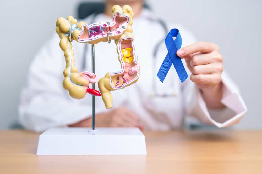
A colonoscopy may identify an abnormal area or growth. Tissue is then collected so a pathologist can determine whether cancer is present. When surgery follows, the resection specimen can provide substantially more information than the initial biopsy.
A clinician examines the inside of the colon and identifies tissue that may require sampling.
A small tissue sample is collected from the abnormal area and sent to the pathology laboratory.
The pathologist examines the biopsy and reports whether cancer is present and what type it appears to be.
If surgery is performed, the removed colon and lymph nodes are examined to provide a more complete pathologic assessment.
These reports answer different questions. A biopsy can confirm that cancer is present, but it usually cannot provide the same level of detail as examination of the surgically removed tumour and surrounding tissue.
A biopsy examines a small portion of the abnormal area. It can often establish the diagnosis and identify the tumour type.
A resection report examines the tumour, bowel wall, surrounding tissue, surgical margins, and regional lymph nodes.
A colon pathology report contains several findings that must be understood together. The amount of information available depends on whether the specimen is a small biopsy or a surgical resection.
Most colon cancers are adenocarcinomas, although the report may describe a more specific histologic type or growth pattern.
The report may describe how closely the tumour resembles normal gland-forming tissue. Grade is one part of the overall assessment.
A resection report may show whether the tumour is within the bowel wall, extends into surrounding tissue, reaches the surface, or involves a nearby structure.
The report may state how many lymph nodes were examined and how many contained tumour.
The report may describe whether tumour reaches a cut edge or circumferential margin. The meaning depends on the specimen and procedure.
Separate tumour deposits may be present in surrounding tissue without being located inside an identifiable lymph node.
The report may document lymphovascular invasion, perineural invasion, tumour budding, perforation, or treatment-related changes.
Mismatch repair, MSI, molecular testing, or additional stains may appear in a later addendum or separate report.
Review the terminology, key findings, limitations, and pending results in your existing colon pathology report during a secure virtual consultation.
Depth of invasion describes how far the tumour extends through the different layers of the colon. This information contributes to the pathologic tumour category, but it is not the complete cancer stage by itself.
The report may describe involvement of the inner layers or muscle of the colon wall.
The tumour may extend through the muscle and into the surrounding fatty tissue.
More extensive disease may reach the outer surface or directly involve an adjacent organ or structure.
Colon resection reports may document additional features that are considered alongside the pathologic stage, molecular findings, imaging, and the wider clinical picture.
This means tumour cells are identified within lymphatic channels or blood vessels. It is separate from finding tumour inside a lymph node.
This describes tumour involving or tracking around nerves. Its significance should be interpreted in the context of the full report.
Tumour budding refers to small groups or individual tumour cells at the invasive edge of the cancer.
Tumour deposits are separate tumour foci in the surrounding tissue and are classified separately from identifiable lymph-node involvement.
Some risk-related information may come from the operative report, imaging, or wider clinical record rather than from microscopic examination alone. Your care team combines these findings with the pathology report.
These tests are often discussed together, but they may answer different questions and may appear in different pathology reports, addenda, or molecular-testing documents.
Immunohistochemistry may examine whether the mismatch repair proteins are retained or lost within the tumour cells.
MSI testing is another way of assessing mismatch repair function. Abnormal results may affect treatment discussions and may lead the care team to consider further inherited-risk assessment.
KRAS, NRAS, and BRAF results may appear in a molecular report or addendum. Their relevance depends on the clinical setting and the wider treatment discussion.
Get a patient-friendly explanation of the biomarker findings already documented in your pathology report and understand which results may still be pending.
A pathology report may answer many important questions, but it may not provide the complete picture on its own.
The diagnosis, tumour type, grade, depth of invasion, lymph-node findings, margins, and additional microscopic features may be documented when the available specimen allows.
Mismatch repair studies, MSI testing, molecular results, additional stains, or outside-slide review may still be in progress after the first report is issued.
Complete staging and treatment decisions may also require imaging, evidence of distant disease, operative findings, laboratory results, and assessment by the treating care team.
A pathology report consultation helps you understand the terminology and findings already documented, so you can approach conversations with your treating team with greater context and more focused questions.
Submit the pathology report and relevant records requested for the consultation.
Discuss the report during a private virtual appointment and ask questions about the documented findings.
Better understand the terminology and prepare questions to discuss with your treating care team.
A report explanation and a formal microscopic second opinion are different services. Setting clear boundaries helps you choose the most appropriate option.
The right option depends on whether you need help understanding the written results or an independent review of the pathology slides and tissue.
These answers provide general educational information. Individual reports vary according to the specimen, procedure, testing, and wider clinical situation.
A colonoscopy biopsy can usually determine whether cancer is present in the sampled tissue and may identify the tumour type and grade. Because the biopsy contains only a small amount of tissue, it usually cannot show the complete depth of invasion, all surgical margins, or the full lymph-node findings.
A surgical resection provides the pathologist with the tumour, bowel wall, surrounding tissue, surgical margins, and regional lymph nodes. This allows a more complete assessment than a small biopsy sample.
A surgical pathology report may provide the pathologic tumour and lymph-node categories, but complete staging may also depend on imaging, evidence of distant spread, operative findings, and other clinical information.
A positive lymph node contains tumour within an identifiable lymph node. Tumour deposits are separate tumour foci in surrounding tissue without an identifiable lymph-node structure. They are related findings but are documented and classified separately.
Lymphovascular invasion means tumour cells are identified within lymphatic channels or blood vessels. Perineural invasion describes tumour involving or tracking around nerves. Neither term is the same as finding tumour in a lymph node.
Mismatch repair immunohistochemistry evaluates proteins involved in repairing DNA. The report may describe the proteins as retained or intact, or show loss or deficiency of one or more proteins. Abnormal tumour findings may lead the treating team to consider additional testing, but they do not by themselves confirm an inherited condition.
Yes. A report consultation can explain the written diagnosis, terminology, depth of invasion, lymph nodes, margins, high-risk features, biomarkers, and pending findings. It does not provide an independent microscopic diagnosis unless a separate slide-review service is ordered.
A second opinion may be more appropriate when the diagnosis is complex, uncertain, unexpected, or when an independent review of the original slides or tissue could provide additional value.
Meet online with a pathologist who can explain the terminology, key findings, biomarker results, and questions raised by your existing report.
Educational support only. This consultation does not replace diagnosis, complete staging, or treatment planning by your treating clinicians.
