
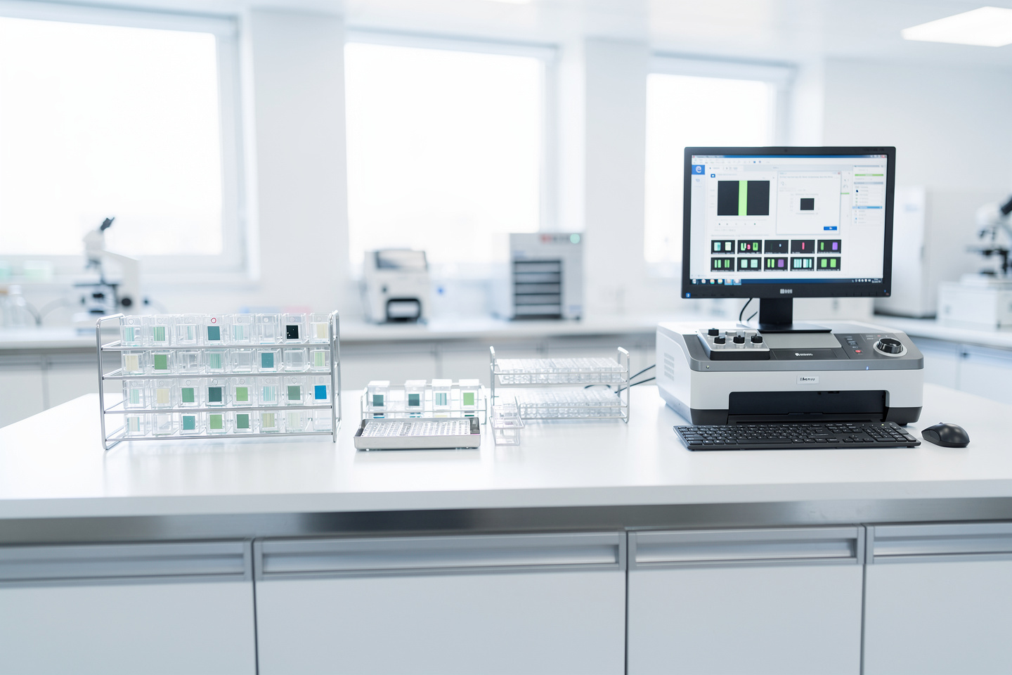
Worried a scan may change your diagnosis? Digital pathology slides are powerful, but here is when glass slides still matter and what to ask.
Seeing the word “digital” in a pathology report can raise understandable questions about how carefully the tissue was reviewed. Digital pathology slides can help pathologists evaluate a case, share it quickly, and support a diagnosis, but they do not always fully replace the original physical glass slides. A digital scan is a high-resolution image made from a thin tissue section on glass. The final answer still depends on the quality of the tissue, the stain, the scan, and the pathologist interpreting what is seen.
Learning that a biopsy or surgical specimen was scanned can feel unsettling for many patients. Common concerns include whether a computer is making the diagnosis, whether something could be missed, or whether a second opinion will be less reliable. In good hands, digital review is a tool, not a shortcut, and it should be used with the same careful medical judgment as microscope review.
What a Scanned Pathology Slide Shows
Digital pathology slides are images created by scanning traditional glass slides at very high resolution. Think of it like turning a paper map into a detailed online map that can be zoomed into, moved around, and shared securely. The map is not the neighborhood itself, but if it is made well, it can show a tremendous amount of useful detail. In pathology, that detail includes cells, tissue patterns, tumor architecture, inflammation, margins, and many special stains.
The physical process starts with a tissue sample from a biopsy or surgery. The laboratory processes that tissue, embeds it in wax, cuts an extremely thin section, places it on glass slides, and stains it so cells become visible. Whole slide imaging then captures the entire slide, usually at magnifications that allow a pathologist to zoom from a broad overview down to individual cells. This is different from a simple photograph through a microscope because the entire slide is scanned in a structured way.
For patients, the key point is that a scanned slide does not erase the role of the pathologist. It changes the way the pathologist views the material. Instead of looking through microscope lenses, the pathologist may review the scanned image on a calibrated medical-grade monitor. A careful microscope review and a careful digital review both require training, context, and correlation with the pathology report, which is explained more fully in What Is a Pathology Report?.
Why a Pathology Case May Be Reviewed Digitally

A report may mention scanned slides, digital review, whole slide imaging, or digital pathology because many hospitals and reference laboratories now use these systems for consultation, archiving, teaching, quality review, and second opinion work. The original glass slides are still made in most routine pathology laboratories because tissue has to be physically cut and stained before it can be scanned. The scan is created after the laboratory work is complete. In that sense, the digital image depends on the quality of the original tissue sample and the physical slide.
Digital pathology is especially helpful when a case needs to be shared with another pathologist quickly. In the past, glass slides often had to be mailed, which could take days and carried a small risk of breakage or loss. With a validated system, a scanned slide can be made available to an expert much faster. That can be useful for rare tumors, complicated margins, unusual inflammatory patterns, and cases where a pathology diagnosis needs subspecialty review.
The lab also has to check whether the scan is technically acceptable. The pathologist looks for focus problems, missing tissue, air bubbles, folds, staining issues, and areas that may need direct microscope review. A digital scan should represent the entire slide, not just a selected field. If the image quality is poor, if the tissue is very thick, or if the stain does not scan well, the pathologist may go back to the glass slides to confirm the finding.
When Glass Slide Review Still Matters
Digital pathology slides are not serious or dangerous by themselves. The more important question is whether the digital method is appropriate for the specific case and whether the laboratory has validated the system for diagnostic use. When the system is properly validated, whole slide imaging can be a safe and efficient way to support a pathology diagnosis. When the system is not validated, or when image quality is inadequate, the pathologist should not rely on the scan alone.
Digital review is usually most reassuring when the scan is sharp, complete, and reviewed by a qualified pathologist who knows when to request the original glass slides. Many routine biopsies, cancer resections, and second opinion cases can be reviewed digitally with excellent accuracy. However, some situations may still call for direct microscope review. Examples include very subtle organisms, certain blood or bone marrow findings, some polarized light evaluations, and cases where the scanned image does not show enough crisp detail.
The word “digital” can feel impersonal because it may bring to mind automation or artificial intelligence. In most patient care settings, the scan is simply the viewing method, while the human pathologist still makes the interpretation. Artificial intelligence tools may assist with counting, measuring, or screening in some settings, but they do not replace the medical responsibility of the pathologist. If a case is complex, or if treatment will change based on the result, asking whether the original glass slides are available for review is reasonable and appropriate.
What Happens After Digital Slide Review
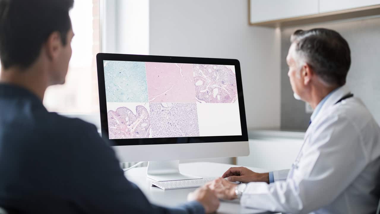
What happens next depends on why the slides were scanned. If the scan was used for routine sign-out, the doctor may simply receive the final pathology report and move forward with the treatment plan. If the scan was used for consultation, a second pathologist may issue an additional opinion or addendum. For patients trying to understand the wording of a report, Understanding Your Pathology Report: How to Read It with Confidence can help connect the technical terms to real clinical decisions.
When treatment depends on the diagnosis, doctors may combine the pathology diagnosis with imaging, blood tests, molecular tests, and the clinical story. Digital pathology can make that process faster because whole slide imaging allows secure sharing between institutions. Still, treatment should not be based on speed alone. The safest approach is careful review, especially when the diagnosis is rare, unexpected, or different from what the treating team anticipated.
When seeking a second opinion, patients can ask whether the reviewing pathologist needs digital files, glass slides, paraffin blocks, or all of these. Some institutions can review digital pathology slides directly, while others still require physical material. If additional stains or molecular tests are needed, the laboratory may need the block, not just the scanned image. For practical next steps, How to Get a Second Opinion on Your Pathology Diagnosis may be helpful.
Questions to Ask About the Slide Review
- Was the diagnosis made from digital pathology slides, glass slides, or both?
- Was the whole slide imaging system validated for diagnostic use in this laboratory?
- Were any parts of the slide blurry, missing, folded, or difficult to evaluate?
- Are the original glass slides and tissue blocks still available if another review is needed?
- Would a microscope review add value for this specific pathology diagnosis?
- Does this case need subspecialty pathology review or a second opinion?
- Could any additional stains, biomarkers, or molecular tests change the interpretation?
These questions are not meant to challenge the care team. They are meant to give patients a clearer picture of how the diagnosis was made. A good pathologist will understand why a patient may want to know whether the diagnosis came from digital review, direct microscope review, or both. A good clinician will also understand that the format of slide review matters when major treatment decisions are being made.
If something about a report does not match what was explained clinically, or if the diagnosis feels surprising, a second opinion can be very helpful. This is especially true for rare tumors, borderline findings, unusual biopsies, or diagnoses that lead to surgery, chemotherapy, radiation, or long-term medication. Patients can read more about timing and warning signs in When Should You Get a Second Pathology Opinion?. The goal is not to create doubt; the goal is to make sure the evidence has been reviewed in the right way.
Frequently Asked Questions
Can digital scans replace physical pathology slides?
In some validated settings, a pathologist can make a reliable diagnosis from a high-quality digital scan. However, the physical material still matters. The tissue section begins on a glass slide, and the tissue block may be needed later for additional stains, biomarkers, or molecular testing. If the scan is incomplete, blurry, or technically limited, the pathologist should review the glass slide or request a better section.
Are digital pathology slides as accurate as glass slides?
They can be highly accurate when the laboratory has properly validated whole slide imaging and the image quality is strong. Accuracy also depends on the specimen type, stain, and diagnostic question. Some findings are well suited to digital review, while others are better confirmed at the microscope. Patients can ask whether anything about the specific case makes glass slide review preferable.
Does a computer make my pathology diagnosis?
In routine medical practice, the diagnosis is made by a pathologist, not by the computer. The computer displays the scanned image and may help with viewing, measuring, or organizing the case. Digital pathology supports the trained physician’s interpretation. If artificial intelligence was used in a special way, patients can ask what it did and whether a pathologist confirmed the result.
Can I get a second opinion from scanned slides?
Yes. Many second opinion reviews can start with scanned slides, particularly when a fast review is important. Requirements vary by institution: some accept digital files, while others need glass slides or tissue blocks before issuing a formal opinion. If the existing slides do not answer the diagnostic question, the reviewing pathologist may request additional stains or other testing.
What if my scanned slide quality was poor?
A poor-quality scan should not be used as the only basis for a difficult interpretation. The laboratory may rescan the slide, clean it, cut a new section, or have the pathologist review the glass slide under the microscope. This is a routine safety step, not necessarily a sign of an error. The goal is to make sure the tissue is clear enough to support an accurate diagnosis.
If a case involves scanned pathology slides, patients are allowed to ask how they were used and whether the original slides remain available. Clarity is part of good care, and patients should not have to guess how a diagnosis was made. Honest Pathology consultations can help patients understand what a report says, what the slide review means, and what questions to bring back to the care team.
References:
FDA — FDA Allows Marketing of First Whole Slide Imaging System for Digital Pathology
College of American Pathologists — Validating Whole Slide Imaging Systems for Diagnostic Purposes in Pathology
NCI Dictionary of Cancer Terms — Biopsy
