When someone is diagnosed with cancer, one of the first questions doctors ask is whether the cancer has spread, especially to the lymph nodes. This detail is incredibly important because it helps determine the stage of cancer, guides treatment decisions, and gives insight into the prognosis.
But how exactly do doctors and pathologists figure that out? Let’s walk through the process in simple, honest terms, and show how Honest Pathology™ helps you understand every detail of what’s written in your report.
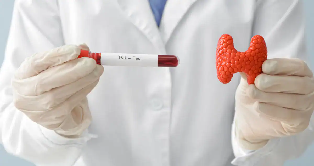
What Are Lymph Nodes, and Why Do They Matter
Lymph nodes are small, bean-shaped glands that are part of your lymphatic system, a network that helps your body fight infection and clear away waste. You have hundreds of lymph nodes all over your body, especially in clusters in your neck, armpits, chest, abdomen, and groin.
When cancer spreads, it often travels first through the lymphatic vessels to the nearest group of lymph nodes. That’s why these nodes are one of the first places doctors check after a cancer diagnosis.
Finding cancer cells in the lymph nodes doesn’t necessarily mean the cancer has gone everywhere , but it does mean it has started to move beyond its original location, which changes how doctors plan your treatment.
How Do Doctors Check the Lymph Nodes
The process usually starts with imaging studies and then, if needed, tissue sampling. Let’s go step by step.
Physical Examination
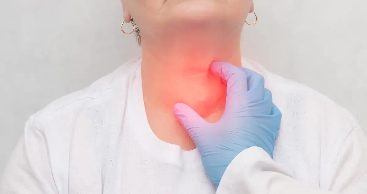
During an exam, your doctor may gently feel areas like your neck, armpits, or groin to see if any lymph nodes feel enlarged or firm.
But not all swollen lymph nodes mean cancer. Infections can cause them to enlarge too. So a physical exam alone can’t confirm cancer spread, it just gives clues that more testing is needed.
Imaging Tests
Doctors often order imaging tests to look at deeper lymph nodes that can’t be felt during an exam. These include:
- CT scans (Computed Tomography) detailed 3-D X-ray images showing the size and shape of lymph nodes.
- MRI (Magnetic Resonance Imaging) provides high-resolution images of soft tissues.
- PET scans (Positron Emission Tomography) highlight areas of increased activity where cancer cells may be growing.
- Ultrasound often used for lymph nodes in the neck or armpit to see their shape and internal texture.
If imaging shows suspicious lymph nodes, doctors may decide to sample them to know for sure.
Needle Biopsy
A needle biopsy is a simple way to collect cells from a lymph node without major surgery. There are two main types:
- Fine Needle Aspiration (FNA): A thin needle collects a small number of cells.
- Core Needle Biopsy: A larger needle removes a small tissue core for a more detailed look.
The sample is sent to the pathology lab, where a pathologist examines it under the microscope to see if any cancer cells are present.
This step is crucial , because even if a node looks enlarged or suspicious on a scan, only a pathologist’s microscopic examination can confirm whether it truly contains cancer.
Sentinel Lymph Node Biopsy
When certain cancers are diagnosed , especially breast cancer, melanoma, and some gynecologic cancers , doctors perform a sentinel lymph node biopsy during surgery.
The “sentinel node” is the first lymph node (or few nodes) that drain lymph fluid from the tumor area , basically, the first stop for any cancer cells that might be trying to spread.
During surgery, the surgeon injects a dye or radioactive tracer near the tumor. This tracer travels to the sentinel node(s), which the surgeon then removes and sends to the pathologist for detailed examination.
If the sentinel nodes are free of cancer, it often means the cancer has not spread further, and additional lymph node removal may not be needed.
If cancer is found there, it suggests a higher likelihood that other nearby nodes could also be affected.
Surgical Lymph Node Dissection
In some cases, especially if the sentinel nodes or needle biopsies are positive, doctors remove a larger cluster of lymph nodes to check how many contain cancer.
This is called a lymph node dissection (or lymphadenectomy). The nodes are then sent to pathology to be examined one by one.
What Happens in the Pathology Lab
Now let’s take a closer look at what the pathologist does , this is where Honest Pathology™ comes in.
1. Gross Examination
The first step is called the gross exam , the pathologist inspects the lymph nodes with the naked eye, noting their number, size, color, and texture. Some nodes may look normal; others might appear enlarged or firm.
Each node is carefully sliced into thin sections and placed into labeled containers for microscopic processing.
2. Microscopic Examination
Under the microscope, the pathologist looks for cancer cells among the normal immune cells of the lymph node.
If cancer cells are seen, the pathologist reports:
- How many lymph nodes were examined
- How many contained cancer
- Whether the cancer deposits are large, small, or microscopic
- If cancer has spread beyond the node capsule (into nearby tissue)
This information goes directly into your pathology report and plays a major role in determining your cancer stage.
3. Special Stains and Immunohistochemistry
Sometimes cancer cells are so small or look very similar to normal cells that they’re hard to detect. In these cases, the pathologist uses special stains or immunohistochemistry (IHC).
IHC involves applying antibodies that bind to specific proteins found on cancer cells, causing them to show up under the microscope in a distinctive color pattern.
This advanced technique helps confirm whether what’s seen is truly cancer , and can even identify the type or origin of cancer cells when it’s not obvious.
4. Micrometastases and Isolated Tumor Cells
Not all lymph node involvement is the same. Pathologists categorize what they find based on size and visibility. Macrometastasis refers to a visible cluster of cancer cells larger than 2 millimeters. Micrometastasis describes tiny clusters between 0.2 and 2 millimeters that can only be seen under a microscope. Isolated tumor cells are scattered single cells or clusters smaller than 0.2 millimeters.
Even these tiny findings can be important, but their significance depends on the type of cancer. Your pathology report will specify which of these are present so your doctor can interpret what it means for your care.
How Lymph Node Results Affect Cancer Staging
One of the main reasons lymph node analysis is so critical is because it directly impacts staging , the system doctors use to describe how advanced a cancer is.
Most cancers are staged using the TNM system:
- T = Tumor size and extent
- N = Lymph node involvement
- M = Metastasis (spread to distant organs)
The “N” part of this system comes entirely from the pathologist’s findings.
For example:
- N0 means no lymph node involvement.
- N1, N2, N3 indicate increasing levels of spread, depending on how many nodes contain cancer and how far they are from the original tumor.
A change from N0 to N1, even if it’s just a single tiny metastasis, can change the stage and therefore the treatment plan.
How Lymph Node Involvement Influences Treatment
Once the pathology report confirms cancer in the lymph nodes, your care team uses that information to tailor your treatment.
Here’s what that might look like:
- Surgery: Additional nodes may be removed if not already done.
- Radiation therapy: Often recommended for regions where cancer cells might remain.
- Chemotherapy or immunotherapy: Used to kill any microscopic cancer cells that could be circulating elsewhere.
- Targeted therapy or hormonal therapy: Based on the specific type of cancer cells identified.
Lymph node results don’t tell the whole story, but they’re a cornerstone in designing the best, most personalized care plan.
What Does “Lymphovascular Invasion” Mean in a Pathology Report?
Sometimes your pathology report might mention lymphovascular invasion (LVI) , meaning that cancer cells were seen inside small lymphatic or blood vessels near the tumor, even if no lymph nodes are positive yet.
LVI suggests the cancer has the potential to spread, even if it hasn’t been detected in a node. It’s an important detail that your oncologist will consider when deciding on further treatment.
Common Questions About Lymph Node Findings
If my lymph nodes are positive, does that mean my cancer is incurable?
Not at all. Many people with lymph node involvement are successfully treated and live long, healthy lives. It simply means your treatment plan may need to be more aggressive or include systemic therapies.
If my lymph nodes are negative, am I “in the clear”?
Negative nodes are a great sign, but your doctor will still monitor you closely. Cancer can sometimes spread in ways that bypass lymph nodes entirely, though that’s less common.
Can imaging alone confirm lymph node spread?
No , imaging can only suggest it. Only a pathologist looking under the microscope can definitively confirm whether cancer cells are in a lymph node.
Why did my report say “micrometastasis detected by immunohistochemistry”?
That means the cancer spread was so small it wasn’t visible on routine stains but was picked up using special antibodies , a very sensitive test. It shows how precise modern pathology has become.
Understanding Your Pathology Report with Honest Pathology™
When you read your pathology report, phrases like “positive for metastasis,” “one of fifteen lymph nodes involved,” or “micrometastasis” can be overwhelming.
That’s where Honest Pathology™ comes in.
Our mission is to help patients truly understand their pathology reports , not in medical jargon, but in plain, compassionate language. During your online educational consultation, a U.S.-trained board-certified pathologist will walk you through your report line by line, explain what the lymph node findings mean, and answer all your questions.
You’ll learn:
- What your lymph node results mean for your stage and treatment options
- How to interpret medical terms like “N1,” “micrometastasis,” or “extracapsular extension”
- What questions to ask your care team next
We never diagnose or treat , but we empower you with knowledge, so you can make informed decisions and approach your next appointment with confidence.
The Honest Pathology™ Difference
When you understand your pathology report, you gain control over your care.
With Honest Pathology™, you’re not just handed a report , you’re guided through it with clarity and compassion.
Because knowledge shouldn’t be a privilege. It should be your right.
Final Thoughts
Knowing whether cancer has spread to the lymph nodes is one of the most important pieces of information in any cancer diagnosis. Through imaging, surgery, and careful examination under the microscope, doctors and pathologists work together to provide a clear, accurate picture of your disease.
And when you’re ready to make sense of that information, Honest Pathology™ is here to help you see the story behind the report , so you can move forward informed, supported, and empowered.